MENU
Bibliography Options Menu
The Electronic Scholarly Publishing Project: Providing world-wide, free access to classic scientific papers and other scholarly materials, since 1993.
More About: ESP | OUR CONTENT | THIS WEBSITE | WHAT'S NEW | WHAT'S HOT
ESP: PubMed Auto Bibliography 30 Jul 2026 at 01:40 Created:
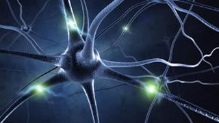
Brain-Computer Interface
Wikipedia: A brain–computer interface (BCI), sometimes called a neural control interface (NCI), mind–machine interface (MMI), direct neural interface (DNI), or brain–machine interface (BMI), is a direct communication pathway between an enhanced or wired brain and an external device. BCIs are often directed at researching, mapping, assisting, augmenting, or repairing human cognitive or sensory-motor functions. Research on BCIs began in the 1970s at the University of California, Los Angeles (UCLA) under a grant from the National Science Foundation, followed by a contract from DARPA. The papers published after this research also mark the first appearance of the expression brain–computer interface in scientific literature. BCI-effected sensory input: Due to the cortical plasticity of the brain, signals from implanted prostheses can, after adaptation, be handled by the brain like natural sensor or effector channels. Following years of animal experimentation, the first neuroprosthetic devices implanted in humans appeared in the mid-1990s. BCI-effected motor output: When artificial intelligence is used to decode neural activity, then send that decoded information to some kind of effector device, BCIs have the potential to restore communication to people who have lost the ability to move or speak. To date, the focus has largely been on motor skills such as reaching or grasping. However, in May of 2021 a study showed that an AI/BCI system could be use to translate thoughts about handwriting into the output of legible characters at a usable rate (90 characters per minute with 94% accuracy).
Created with PubMed® Query: (bci OR (brain-computer OR brain-machine OR mind-machine OR neural-control interface) NOT 26799652[PMID] ) NOT pmcbook NOT ispreviousversion
Citations The Papers (from PubMed®)
RevDate: 2026-07-29
CmpDate: 2026-07-29
Correction: Modeling the acceptability of BCIs for motor rehabilitation after stroke: a large scale study on the general public.
Frontiers in neuroergonomics, 7:1854498.
[This corrects the article DOI: 10.3389/fnrgo.2022.1082901.].
Additional Links: PMID-42519170
Full Text:
Publisher:
PubMed:
Citation:
show bibtex listing
hide bibtex listing
@article {pmid42519170,
year = {2026},
author = {Grevet, E and Forge, K and Tadiello, S and Izac, M and Amadieu, F and Brunel, L and Pillette, L and Py, J and Gasq, D and Jeunet-Kelway, C},
title = {Correction: Modeling the acceptability of BCIs for motor rehabilitation after stroke: a large scale study on the general public.},
journal = {Frontiers in neuroergonomics},
volume = {7},
number = {},
pages = {1854498},
doi = {10.3389/fnrgo.2026.1854498},
pmid = {42519170},
issn = {2673-6195},
abstract = {[This corrects the article DOI: 10.3389/fnrgo.2022.1082901.].},
}
RevDate: 2026-07-29
CmpDate: 2026-07-29
Correction: Domain-aware domain-class adaptation network for motor execution to motor imagery EEG classification.
Frontiers in neuroscience, 20:1903945.
[This corrects the article DOI: 10.3389/fnins.2026.1851006.].
Additional Links: PMID-42519235
Full Text:
Publisher:
PubMed:
Citation:
show bibtex listing
hide bibtex listing
@article {pmid42519235,
year = {2026},
author = {Wang, J and Xu, G and Du, C and Li, Z and Li, H and Chen, S and Han, C and Zhang, S},
title = {Correction: Domain-aware domain-class adaptation network for motor execution to motor imagery EEG classification.},
journal = {Frontiers in neuroscience},
volume = {20},
number = {},
pages = {1903945},
doi = {10.3389/fnins.2026.1903945},
pmid = {42519235},
issn = {1662-4548},
abstract = {[This corrects the article DOI: 10.3389/fnins.2026.1851006.].},
}
RevDate: 2026-07-28
A glutamatergic S1-VPL-S1 corticothalamocortical loop amplifies mechanical hypersensitivity in neuropathic pain.
Pain [Epub ahead of print].
Although cortical-thalamic projections contribute to pain modulation, their downstream targets and functional roles are not well defined. Here, we show that glutamatergic neurons in the hindlimb region of primary somatosensory cortex (S1HLGlu) project to a subset of glutamatergic neurons in the ventral posterolateral thalamic nucleus (VPLGlu) that are anatomically separated from VPL neurons receiving peripheral afferent inputs. These VPLGlu neurons preferentially innervate S1HLGlu neurons, forming a recurrent glutamatergic corticothalamocortical pathway. Optogenetic and chemogenetic activation of the S1HLGlu-VPLGlu-S1HLGlu pathway reduced mechanical withdrawal thresholds under baseline conditions, whereas circuit inhibition alleviated mechanical allodynia and spontaneous pain in the spared nerve injury model without altering baseline nociception of mice. In vivo fiber photometry further demonstrated that this pathway enhanced S1HLGlu responses to normally subthreshold mechanical stimuli, suggesting a role in sensory amplification. Together, these findings indicate that the S1HLGlu-VPLGlu-S1HLGlu pathway becomes pathologically engaged during neuropathic pain and contributes to mechanical hypersensitivity. This work provides mechanistic insight into corticothalamic involvement in cortical pain processing and highlights a selective excitatory pathway as a potential target for neuropathic pain intervention.
Additional Links: PMID-42520165
PubMed:
Citation:
show bibtex listing
hide bibtex listing
@article {pmid42520165,
year = {2026},
author = {Hu, Y and Ding, H and Zou, L and Wen, Z and Wu, H and Ni, T and He, L and Gao, Q and Cao, H and Zhou, J and Chen, J and Jiang, B and Qiu, S and Yu, L and Yan, M},
title = {A glutamatergic S1-VPL-S1 corticothalamocortical loop amplifies mechanical hypersensitivity in neuropathic pain.},
journal = {Pain},
volume = {},
number = {},
pages = {},
pmid = {42520165},
issn = {1872-6623},
support = {82371217//the National Natural Science Foundation of China/ ; 82300566//National Natural Science Foundation of China/ ; 82071227//National Natural Science Foundation of China/ ; LZ23H090003//Natural Science Foundation of Zhejiang Province/ ; No.18 (2020)//the Leading Health Talents of Zhejiang Province, Zhejiang Health Office/ ; 2021-595 LCZDZK-01//the National Clinical Key Specialty Construction Project of China/ ; 2023M733095//the China Postdoctoral Science Foundation/ ; 2025T180562//the China Postdoctoral Science Foundation/ ; 82471230//National Natural Science Foundation of China/ ; },
abstract = {Although cortical-thalamic projections contribute to pain modulation, their downstream targets and functional roles are not well defined. Here, we show that glutamatergic neurons in the hindlimb region of primary somatosensory cortex (S1HLGlu) project to a subset of glutamatergic neurons in the ventral posterolateral thalamic nucleus (VPLGlu) that are anatomically separated from VPL neurons receiving peripheral afferent inputs. These VPLGlu neurons preferentially innervate S1HLGlu neurons, forming a recurrent glutamatergic corticothalamocortical pathway. Optogenetic and chemogenetic activation of the S1HLGlu-VPLGlu-S1HLGlu pathway reduced mechanical withdrawal thresholds under baseline conditions, whereas circuit inhibition alleviated mechanical allodynia and spontaneous pain in the spared nerve injury model without altering baseline nociception of mice. In vivo fiber photometry further demonstrated that this pathway enhanced S1HLGlu responses to normally subthreshold mechanical stimuli, suggesting a role in sensory amplification. Together, these findings indicate that the S1HLGlu-VPLGlu-S1HLGlu pathway becomes pathologically engaged during neuropathic pain and contributes to mechanical hypersensitivity. This work provides mechanistic insight into corticothalamic involvement in cortical pain processing and highlights a selective excitatory pathway as a potential target for neuropathic pain intervention.},
}
RevDate: 2026-07-28
Combined effects of an ointment comprising Eisenia fetida oil, Valeriana officinalis, and zinc oxide nanoparticles on healing in a rat model of second-degree burns.
Burns : journal of the International Society for Burn Injuries, 52(8):108140 pii:S0305-4179(26)00292-5 [Epub ahead of print].
Burns are one of the most devastating traumatic injuries, and in cases of advanced burns, patients require urgent and specialized care to minimize mortality. Studies have shown that a variety of herbal medicines and nanoparticles can be used to treat burn wounds. Among them mixture of Eisenia fetida oil, valerian, and zinc oxide (ZnO) nanoparticles can be a good choice due to their healing properties. This study aimed to synthesize an ointment based on Eisenia fetida oil, Valeriana officinalis, and zinc oxide to investigate its effect on skin wound healing. The hemocompatibility and biocompatibility of the designed ointment were evaluated using blood clotting index (BCI), hemolysis test, and 3-(4,5-dimethylthiazol-2-yl)-2,5-diphenyl tetrazolium bromide (MTT) test. The antibacterial effects of the designed ointment were evaluated using minimum inhibitory concentration (MIC), minimum bactericidal concentration (MBC), and time-kill tests. Finally, the healing ability of the designed ointment was evaluated in a rat model by creating a 3 × 3 cm burn wound. Wound healing was evaluated after 14 days by hematoxylin-eosin (H&E) and Verhoeff-Van Gieson (VVG) staining. The results showed that the addition of Valeriana officinalis and zinc oxide to Eisenia fetida oil improved hemocompatibility, biocompatibility, improved cell migration, and improved antibacterial properties. Additionally, the results of H&E and VVG staining demonstrated the good efficacy of the designed ointment in increasing epidermal thickness, promoting angiogenesis, and enhancing collagen and elastin fibers. These results indicate the potential of the designed ointment as a promising tool for skin wound healing and clinical trials.
Additional Links: PMID-42520418
Publisher:
PubMed:
Citation:
show bibtex listing
hide bibtex listing
@article {pmid42520418,
year = {2026},
author = {Tabatabai, TS and Tabatabai, TS and Vaez, A and Ehterami, A and Atashi, A and Semyari, H and Salehi, M},
title = {Combined effects of an ointment comprising Eisenia fetida oil, Valeriana officinalis, and zinc oxide nanoparticles on healing in a rat model of second-degree burns.},
journal = {Burns : journal of the International Society for Burn Injuries},
volume = {52},
number = {8},
pages = {108140},
doi = {10.1016/j.burns.2026.108140},
pmid = {42520418},
issn = {1879-1409},
abstract = {Burns are one of the most devastating traumatic injuries, and in cases of advanced burns, patients require urgent and specialized care to minimize mortality. Studies have shown that a variety of herbal medicines and nanoparticles can be used to treat burn wounds. Among them mixture of Eisenia fetida oil, valerian, and zinc oxide (ZnO) nanoparticles can be a good choice due to their healing properties. This study aimed to synthesize an ointment based on Eisenia fetida oil, Valeriana officinalis, and zinc oxide to investigate its effect on skin wound healing. The hemocompatibility and biocompatibility of the designed ointment were evaluated using blood clotting index (BCI), hemolysis test, and 3-(4,5-dimethylthiazol-2-yl)-2,5-diphenyl tetrazolium bromide (MTT) test. The antibacterial effects of the designed ointment were evaluated using minimum inhibitory concentration (MIC), minimum bactericidal concentration (MBC), and time-kill tests. Finally, the healing ability of the designed ointment was evaluated in a rat model by creating a 3 × 3 cm burn wound. Wound healing was evaluated after 14 days by hematoxylin-eosin (H&E) and Verhoeff-Van Gieson (VVG) staining. The results showed that the addition of Valeriana officinalis and zinc oxide to Eisenia fetida oil improved hemocompatibility, biocompatibility, improved cell migration, and improved antibacterial properties. Additionally, the results of H&E and VVG staining demonstrated the good efficacy of the designed ointment in increasing epidermal thickness, promoting angiogenesis, and enhancing collagen and elastin fibers. These results indicate the potential of the designed ointment as a promising tool for skin wound healing and clinical trials.},
}
RevDate: 2026-07-28
Distinct neural modes carry information about attempted grasp timing and force in the sensorimotor cortex.
The Journal of neuroscience : the official journal of the Society for Neuroscience pii:JNEUROSCI.0205-26.2026 [Epub ahead of print].
Humans perform a variety of complex hand movements to manipulate objects, requiring precise control of changing forces. Understanding the role of sensorimotor cortex and the cortical dynamics underlying these actions is crucial for developing interventions that restore dexterous hand function after injury or disease. In this study, two male individuals with tetraplegia resulting from cervical spinal cord injury attempted a series of isometric grasps. Neural activity was recorded from the motor and somatosensory cortices using intracortical microelectrode arrays while participants attempted to exert a static or ramping force up and down. Despite their inability to execute movement and limited afferent input, the spiking activity in motor and somatosensory cortex was modulated with the task. Within the neural response we identified independent neural modes - distinct patterns of population-level neural activity that were informative about both the timing and magnitude of the attempted force. Moreover, distinct neural modes were observed during static and dynamic grasping conditions, suggesting independent control schemes for maintaining and changing forces. These modes were related to phases of the task, including the onset, offset, holding periods, as well as increasing and decreasing attempted forces. These results will inform the design of intracortical brain-computer interface (iBCI) systems that can leverage the patterns of grasp and force control evident in sensorimotor cortex during attempted movement to restore dexterous hand function.Significance Statement Restoring dexterous hand function after injury remains a major challenge, partly due to an incomplete understanding of the cortical dynamics underlying grasping and force control. In this study, we investigated neural activity within the motor and somatosensory cortices of individuals with tetraplegia attempting to perform grasps to different target forces with varying temporal profiles. We identified distinct neural modes modulated during specific phases of grasp that encode attempted force information throughout the task. These findings suggest that brain-computer interfaces could leverage these neural modes to restore grasping and force modulation.
Additional Links: PMID-42521627
Publisher:
PubMed:
Citation:
show bibtex listing
hide bibtex listing
@article {pmid42521627,
year = {2026},
author = {Blumenthal, GH and Dekleva, BM and Gontier, C and Gonzalez, IC and Gonzalez-Martinez, JA and Yu, BM and Batista, AP and Sobinov, AR and Miller, LE and Gaunt, RA and Boninger, ML and Chase, SM and Collinger, JL},
title = {Distinct neural modes carry information about attempted grasp timing and force in the sensorimotor cortex.},
journal = {The Journal of neuroscience : the official journal of the Society for Neuroscience},
volume = {},
number = {},
pages = {},
doi = {10.1523/JNEUROSCI.0205-26.2026},
pmid = {42521627},
issn = {1529-2401},
abstract = {Humans perform a variety of complex hand movements to manipulate objects, requiring precise control of changing forces. Understanding the role of sensorimotor cortex and the cortical dynamics underlying these actions is crucial for developing interventions that restore dexterous hand function after injury or disease. In this study, two male individuals with tetraplegia resulting from cervical spinal cord injury attempted a series of isometric grasps. Neural activity was recorded from the motor and somatosensory cortices using intracortical microelectrode arrays while participants attempted to exert a static or ramping force up and down. Despite their inability to execute movement and limited afferent input, the spiking activity in motor and somatosensory cortex was modulated with the task. Within the neural response we identified independent neural modes - distinct patterns of population-level neural activity that were informative about both the timing and magnitude of the attempted force. Moreover, distinct neural modes were observed during static and dynamic grasping conditions, suggesting independent control schemes for maintaining and changing forces. These modes were related to phases of the task, including the onset, offset, holding periods, as well as increasing and decreasing attempted forces. These results will inform the design of intracortical brain-computer interface (iBCI) systems that can leverage the patterns of grasp and force control evident in sensorimotor cortex during attempted movement to restore dexterous hand function.Significance Statement Restoring dexterous hand function after injury remains a major challenge, partly due to an incomplete understanding of the cortical dynamics underlying grasping and force control. In this study, we investigated neural activity within the motor and somatosensory cortices of individuals with tetraplegia attempting to perform grasps to different target forces with varying temporal profiles. We identified distinct neural modes modulated during specific phases of grasp that encode attempted force information throughout the task. These findings suggest that brain-computer interfaces could leverage these neural modes to restore grasping and force modulation.},
}
RevDate: 2026-07-29
CmpDate: 2026-07-29
From behavioral profiles to neural scaffolds: cortico-striatal integrity predicts longitudinal stability and transition in psychological adaptability during early adulthood.
Psychological medicine, 56:e243 pii:S0033291726105352.
BACKGROUND: Psychological adaptability hinges on the dynamic balance between cognitive regulation (self-control) and emotional reactivity (impulsivity, reward/punishment sensitivity). However, traditional variable-centered approaches often fail to capture how these traits holistically co-occur, and the neural architectures predicting their longitudinal transitions in early adulthood remain under-explored.
METHODS: This longitudinal study (N = 1,229 baseline; N = 432 2-year follow-up) integrated a person-centered behavioral approach with resting-state fMRI. We employed Latent Profile Analysis (LPA) to identify trait configurations based on self-control, impulsivity, and sensitivity to reward/punishment. Network-Based Statistic (NBS) analysis with family-wise error (FWE) correction was utilized to evaluate functional connectivity differences among groups. Finally, hierarchical regression and formal interaction models were conducted to test the prospective predictive validity of the identified neural circuits.
RESULTS: LPA delineated three distinct baseline profiles: Adaptive, Moderate, and Maladaptive. NBS analysis revealed that the 'Adaptive' profile is underpinned by robust cortico-striatal functional connectivity, integrating the medial prefrontal cortex and striatum. Longitudinally, initial regression models demonstrated that the baseline integrity of this circuit prospectively predicted behavioral adaptation at the 2-year follow-up. Furthermore, hierarchical regression and formal interaction analyses confirmed that this cortico-striatal circuit provided significant incremental predictive validity - above and beyond massive baseline behavioral stability - specifically in males.
CONCLUSIONS: These findings highlight cortico-striatal integration as a significant, gender-specific prospective predictor of longitudinal adaptation. By bridging person-centered profiling with network neuroscience, this study elucidates the neural correlates supporting future adaptive functioning and psychological resilience during the early adult transition.
Additional Links: PMID-42522230
Publisher:
PubMed:
Citation:
show bibtex listing
hide bibtex listing
@article {pmid42522230,
year = {2026},
author = {He, J and Zhao, H and Lei, X and Qiu, J and Chen, L and Feng, T and Chen, H and Turel, O and He, Q},
title = {From behavioral profiles to neural scaffolds: cortico-striatal integrity predicts longitudinal stability and transition in psychological adaptability during early adulthood.},
journal = {Psychological medicine},
volume = {56},
number = {},
pages = {e243},
doi = {10.1017/S0033291726105352},
pmid = {42522230},
issn = {1469-8978},
support = {31972906//National Natural Science Foundation of China/ ; },
mesh = {Humans ; Magnetic Resonance Imaging ; Longitudinal Studies ; Male ; *Adaptation, Psychological/physiology ; Female ; Adult ; *Corpus Striatum/physiology/diagnostic imaging ; Young Adult ; Impulsive Behavior/physiology ; *Prefrontal Cortex/physiology/diagnostic imaging ; Connectome ; Reward ; },
abstract = {BACKGROUND: Psychological adaptability hinges on the dynamic balance between cognitive regulation (self-control) and emotional reactivity (impulsivity, reward/punishment sensitivity). However, traditional variable-centered approaches often fail to capture how these traits holistically co-occur, and the neural architectures predicting their longitudinal transitions in early adulthood remain under-explored.
METHODS: This longitudinal study (N = 1,229 baseline; N = 432 2-year follow-up) integrated a person-centered behavioral approach with resting-state fMRI. We employed Latent Profile Analysis (LPA) to identify trait configurations based on self-control, impulsivity, and sensitivity to reward/punishment. Network-Based Statistic (NBS) analysis with family-wise error (FWE) correction was utilized to evaluate functional connectivity differences among groups. Finally, hierarchical regression and formal interaction models were conducted to test the prospective predictive validity of the identified neural circuits.
RESULTS: LPA delineated three distinct baseline profiles: Adaptive, Moderate, and Maladaptive. NBS analysis revealed that the 'Adaptive' profile is underpinned by robust cortico-striatal functional connectivity, integrating the medial prefrontal cortex and striatum. Longitudinally, initial regression models demonstrated that the baseline integrity of this circuit prospectively predicted behavioral adaptation at the 2-year follow-up. Furthermore, hierarchical regression and formal interaction analyses confirmed that this cortico-striatal circuit provided significant incremental predictive validity - above and beyond massive baseline behavioral stability - specifically in males.
CONCLUSIONS: These findings highlight cortico-striatal integration as a significant, gender-specific prospective predictor of longitudinal adaptation. By bridging person-centered profiling with network neuroscience, this study elucidates the neural correlates supporting future adaptive functioning and psychological resilience during the early adult transition.},
}
MeSH Terms:
show MeSH Terms
hide MeSH Terms
Humans
Magnetic Resonance Imaging
Longitudinal Studies
Male
*Adaptation, Psychological/physiology
Female
Adult
*Corpus Striatum/physiology/diagnostic imaging
Young Adult
Impulsive Behavior/physiology
*Prefrontal Cortex/physiology/diagnostic imaging
Connectome
Reward
RevDate: 2026-07-29
CmpDate: 2026-07-29
When Music Loses Its Pleasure: Hippocampal Cingulum White Matter as a Structural Mediator of Musical Reward Decline in Aging.
bioRxiv : the preprint server for biology pii:2026.07.16.738989.
UNLABELLED: Individuals' ability to obtain pleasure from music, referred to as musical reward sensitivity, declines with age, yet the neural mechanisms underlying this decline remain unclear. In this study, we investigated musical reward sensitivity measured in 58 older adults and 131 young adults. Consistent with prior findings, young adults reported higher musical reward sensitivity than older adults (p < 0.05). To identify neuroanatomical predictors of musical reward sensitivity, we employed the elastic-net model to predict musical reward sensitivity using white matter microstructural properties and gray matter morphometric properties from the whole brain. In older adults, fractional anisotropy (FA) in the bilateral hippocampal cingulum (CGH) and external capsule (EC) reliably predicted individual differences in musical reward sensitivity. Moreover, FA in the right CGH significantly mediated the relationship between age and musical reward sensitivity in older adults. Notably, these associations were specific to musical reward sensitivity in older adults-that is, they did not replicate in young adults, and did not extend to general reward sensitivity. Together, these findings highlight the critical role of white matter integrity, particularly within the hippocampal-limbic pathways, in age-related changes in musical reward processing, and suggest a potential neurobiological target for interventions aimed at enhancing well-being in older adulthood.
KEY POINTS: Whole-brain machine learning identified the FA in the hippocampal cingulum and external capsule as robust predictors of musical reward sensitivity in older adults.FA in the right hippocampal cingulum mediated the association between age and musical reward sensitivity.These findings extend previous association-based studies by identifying hippocampal-limbic white matter as a key neural substrate supporting musical reward sensitivity decline during aging.
Additional Links: PMID-42523358
Full Text:
Publisher:
PubMed:
Citation:
show bibtex listing
hide bibtex listing
@article {pmid42523358,
year = {2026},
author = {Wang, J and Kathios, N and Kubit, B and Lopez, K and Kim, JC and Large, E and Noble, S and Loui, P},
title = {When Music Loses Its Pleasure: Hippocampal Cingulum White Matter as a Structural Mediator of Musical Reward Decline in Aging.},
journal = {bioRxiv : the preprint server for biology},
volume = {},
number = {},
pages = {},
doi = {10.64898/2026.07.16.738989},
pmid = {42523358},
issn = {2692-8205},
abstract = {UNLABELLED: Individuals' ability to obtain pleasure from music, referred to as musical reward sensitivity, declines with age, yet the neural mechanisms underlying this decline remain unclear. In this study, we investigated musical reward sensitivity measured in 58 older adults and 131 young adults. Consistent with prior findings, young adults reported higher musical reward sensitivity than older adults (p < 0.05). To identify neuroanatomical predictors of musical reward sensitivity, we employed the elastic-net model to predict musical reward sensitivity using white matter microstructural properties and gray matter morphometric properties from the whole brain. In older adults, fractional anisotropy (FA) in the bilateral hippocampal cingulum (CGH) and external capsule (EC) reliably predicted individual differences in musical reward sensitivity. Moreover, FA in the right CGH significantly mediated the relationship between age and musical reward sensitivity in older adults. Notably, these associations were specific to musical reward sensitivity in older adults-that is, they did not replicate in young adults, and did not extend to general reward sensitivity. Together, these findings highlight the critical role of white matter integrity, particularly within the hippocampal-limbic pathways, in age-related changes in musical reward processing, and suggest a potential neurobiological target for interventions aimed at enhancing well-being in older adulthood.
KEY POINTS: Whole-brain machine learning identified the FA in the hippocampal cingulum and external capsule as robust predictors of musical reward sensitivity in older adults.FA in the right hippocampal cingulum mediated the association between age and musical reward sensitivity.These findings extend previous association-based studies by identifying hippocampal-limbic white matter as a key neural substrate supporting musical reward sensitivity decline during aging.},
}
RevDate: 2026-07-29
CmpDate: 2026-07-29
Decoding and Characterizing the Intracranial Representation of Semantic Information.
bioRxiv : the preprint server for biology pii:2026.07.13.738249.
Brain-computer interfaces (BCIs) have achieved impressive performance by decoding motor and articulatory signals associated with speech production. However, considerably less is known about whether higher-level semantic representations can be decoded from human cortical activity. Demonstrating semantic decoding would advance both our understanding of language organization and the development of BCIs that rely on conceptual rather than purely articulatory information. We recorded intracranial neural activity from patients undergoing stereotactic electroencephalography (sEEG) for clinical epilepsy monitoring while they performed language tasks requiring semantic processing. High-gamma power was extracted from local field potentials and used to generate trial-level features for supervised machine-learning classification. Classification performance was evaluated using cross-validation. Semantic category information was decoded significantly above chance, with mean classification accuracy reaching 29.8% across 15 semantic categories (chance = 6.7%). These findings demonstrate that high-gamma activity contains information about conceptual category membership that can be extracted on individual trials. These results provide evidence that semantic information is accessible from intracranial population recordings and support the feasibility of semantic decoding as a complementary direction for future language BCIs. Beyond neuroprosthetic applications, this work contributes to understanding how conceptual knowledge is represented in the distributed human language network.
Additional Links: PMID-42523363
Full Text:
Publisher:
PubMed:
Citation:
show bibtex listing
hide bibtex listing
@article {pmid42523363,
year = {2026},
author = {Smith, C and Inchyna, S and Barrentine, B and Nelson, M},
title = {Decoding and Characterizing the Intracranial Representation of Semantic Information.},
journal = {bioRxiv : the preprint server for biology},
volume = {},
number = {},
pages = {},
doi = {10.64898/2026.07.13.738249},
pmid = {42523363},
issn = {2692-8205},
abstract = {Brain-computer interfaces (BCIs) have achieved impressive performance by decoding motor and articulatory signals associated with speech production. However, considerably less is known about whether higher-level semantic representations can be decoded from human cortical activity. Demonstrating semantic decoding would advance both our understanding of language organization and the development of BCIs that rely on conceptual rather than purely articulatory information. We recorded intracranial neural activity from patients undergoing stereotactic electroencephalography (sEEG) for clinical epilepsy monitoring while they performed language tasks requiring semantic processing. High-gamma power was extracted from local field potentials and used to generate trial-level features for supervised machine-learning classification. Classification performance was evaluated using cross-validation. Semantic category information was decoded significantly above chance, with mean classification accuracy reaching 29.8% across 15 semantic categories (chance = 6.7%). These findings demonstrate that high-gamma activity contains information about conceptual category membership that can be extracted on individual trials. These results provide evidence that semantic information is accessible from intracranial population recordings and support the feasibility of semantic decoding as a complementary direction for future language BCIs. Beyond neuroprosthetic applications, this work contributes to understanding how conceptual knowledge is represented in the distributed human language network.},
}
RevDate: 2026-07-29
CmpDate: 2026-07-29
Dynamic frailty and depressive symptoms in relation to incident stroke: findings from five harmonized longitudinal cohorts.
Frontiers in neurology, 17:1880619.
BACKGROUND: Frailty and depressive symptoms are common in later life and may be related to cerebrovascular risk. Evidence remains limited on whether frailty burden and frailty change are associated with incident stroke across diverse aging cohorts.
METHODS: We analyzed harmonized longitudinal data from five population-based aging cohorts: the Health and Retirement Study (HRS), China Health and Retirement Longitudinal Study (CHARLS), Survey of Health, Ageing and Retirement in Europe (SHARE), English Longitudinal Study of Ageing (ELSA), and Mexican Health and Aging Study (MHAS). Frailty was measured using a harmonized 24-item deficit-accumulation frailty index (FI). The primary analysis used cohort-specific Cox proportional hazards models to estimate associations between baseline FI and first observed incident stroke during follow-up. Secondary exploratory analyses evaluated nonlinearity, FI change, competing mortality, depressive symptoms as a pathway marker, and two-wave cross-lagged associations.
RESULTS: The analytic sample included 81482 participants and 5,089 incident stroke events. In fully adjusted cohort-specific Cox models, each 0.1-unit increase in FI was associated with higher stroke risk in HRS, CHARLS, SHARE, and MHAS, but not in ELSA. Substantial between-cohort heterogeneity was observed; therefore, cohort-specific estimates were interpreted as the primary results and the random-effects pooled estimate was treated as descriptive. Fine-Gray sensitivity analyses treating death as a competing event supported positive frailty-stroke associations across all five cohorts. Restricted cubic spline (RCS) analyses suggested nonlinear associations for baseline FI and FI change. Exploratory pathway analyses indicated that depressive symptoms statistically accounted for part of selected frailty-stroke associations, although patterns varied by cohort and exposure definition. Two-wave cross-lagged panel models (CLPMs) suggested small, cohort-specific prospective associations between elevated frailty vulnerability and later depressive symptoms or stroke; these findings were interpreted as exploratory temporal associations rather than causal within-person effects.
CONCLUSION: Higher frailty burden was associated with incident stroke in most, but not all, harmonized aging cohorts, with substantial heterogeneity across populations. The findings support repeated frailty assessment and integrated mood evaluation in older adults while emphasizing the need for cohort-specific interpretation and confirmatory studies with adjudicated stroke outcomes.
Additional Links: PMID-42523883
PubMed:
Citation:
show bibtex listing
hide bibtex listing
@article {pmid42523883,
year = {2026},
author = {Jiang, Y and Wang, Q and Zhang, J and Shan, M and Guo, Q},
title = {Dynamic frailty and depressive symptoms in relation to incident stroke: findings from five harmonized longitudinal cohorts.},
journal = {Frontiers in neurology},
volume = {17},
number = {},
pages = {1880619},
pmid = {42523883},
issn = {1664-2295},
abstract = {BACKGROUND: Frailty and depressive symptoms are common in later life and may be related to cerebrovascular risk. Evidence remains limited on whether frailty burden and frailty change are associated with incident stroke across diverse aging cohorts.
METHODS: We analyzed harmonized longitudinal data from five population-based aging cohorts: the Health and Retirement Study (HRS), China Health and Retirement Longitudinal Study (CHARLS), Survey of Health, Ageing and Retirement in Europe (SHARE), English Longitudinal Study of Ageing (ELSA), and Mexican Health and Aging Study (MHAS). Frailty was measured using a harmonized 24-item deficit-accumulation frailty index (FI). The primary analysis used cohort-specific Cox proportional hazards models to estimate associations between baseline FI and first observed incident stroke during follow-up. Secondary exploratory analyses evaluated nonlinearity, FI change, competing mortality, depressive symptoms as a pathway marker, and two-wave cross-lagged associations.
RESULTS: The analytic sample included 81482 participants and 5,089 incident stroke events. In fully adjusted cohort-specific Cox models, each 0.1-unit increase in FI was associated with higher stroke risk in HRS, CHARLS, SHARE, and MHAS, but not in ELSA. Substantial between-cohort heterogeneity was observed; therefore, cohort-specific estimates were interpreted as the primary results and the random-effects pooled estimate was treated as descriptive. Fine-Gray sensitivity analyses treating death as a competing event supported positive frailty-stroke associations across all five cohorts. Restricted cubic spline (RCS) analyses suggested nonlinear associations for baseline FI and FI change. Exploratory pathway analyses indicated that depressive symptoms statistically accounted for part of selected frailty-stroke associations, although patterns varied by cohort and exposure definition. Two-wave cross-lagged panel models (CLPMs) suggested small, cohort-specific prospective associations between elevated frailty vulnerability and later depressive symptoms or stroke; these findings were interpreted as exploratory temporal associations rather than causal within-person effects.
CONCLUSION: Higher frailty burden was associated with incident stroke in most, but not all, harmonized aging cohorts, with substantial heterogeneity across populations. The findings support repeated frailty assessment and integrated mood evaluation in older adults while emphasizing the need for cohort-specific interpretation and confirmatory studies with adjudicated stroke outcomes.},
}
RevDate: 2026-07-29
Single-unit Characterization of Electrically Evoked Peripheral Nerve Entrainment Failure.
Neuromodulation : journal of the International Neuromodulation Society pii:S1094-7159(26)00630-6 [Epub ahead of print].
OBJECTIVES: Evidence suggests that peripheral somatosensory nerves do not reliably entrain to electric stimulation even at low frequencies (<100 Hz). This is a concern for peripheral neuromodulation devices because it may cause discrepancies between expected and actual stimulation outcomes. To study this, we investigated the relationship between single-unit peripheral nerve responses (spikes) and the duration, frequency, and amplitude of electric stimulation.
MATERIALS AND METHODS: Single-unit teased-fiber recordings of mechanosensitive units were obtained from Sprague Dawley rat sciatic nerves. To characterize spike entrainment failure, electric stimulation was applied to the hindpaw at various durations, frequencies, and amplitudes. In addition, interleaved trains of electric and mechanic stimulus pulses were delivered to examine their interaction in generating responses. Spike response probability, spike latency, and spike amplitude were compared across stimulation conditions using linear mixed-effects regression models. To assess the ability of a standard nerve model to explain our findings, electric stimulation was simulated in a COMSOL/NEURON McIntyre-Richardson-Grill model of myelinated fibers.
RESULTS: Sustained electric stimulation resulted in spike entrainment failure at frequencies as low as 50 Hz. Units were not uniformly affected; those with faster initial conduction velocity were more strongly affected by long-duration (five minutes) electric stimulation at 50 Hz, while slower units displayed more entrainment failure at high frequencies over shorter durations (three seconds). Increased stimulation amplitude restored entrainment. Furthermore, electric stimulation generated changes in both spike latency and amplitude and interfered with mechanically evoked activity. The standard nerve model also showed entrainment failure, but the time course was dissimilar to our in vivo observations.
CONCLUSIONS: Our data support accounts of spike entrainment failure due to electric stimulation in the peripheral somatosensory system. We hypothesize from our findings that initial failure of spike entrainment is related to previously reported slow axonal K[+] channel activity, but progressive failure is better explained by the sodium-potassium pump conductance. This work provides important insight into mechanisms limiting the efficacy of clinical neuromodulation devices.
Additional Links: PMID-42524812
Publisher:
PubMed:
Citation:
show bibtex listing
hide bibtex listing
@article {pmid42524812,
year = {2026},
author = {Su, TF and Qin, P and So, A and Devecioğlu, İ and Vickery, RM and Birznieks, I and Shivdasani, MN and Moalem-Taylor, G and Guo, T and Aplin, FP},
title = {Single-unit Characterization of Electrically Evoked Peripheral Nerve Entrainment Failure.},
journal = {Neuromodulation : journal of the International Neuromodulation Society},
volume = {},
number = {},
pages = {},
doi = {10.1016/j.neurom.2026.06.469},
pmid = {42524812},
issn = {1525-1403},
abstract = {OBJECTIVES: Evidence suggests that peripheral somatosensory nerves do not reliably entrain to electric stimulation even at low frequencies (<100 Hz). This is a concern for peripheral neuromodulation devices because it may cause discrepancies between expected and actual stimulation outcomes. To study this, we investigated the relationship between single-unit peripheral nerve responses (spikes) and the duration, frequency, and amplitude of electric stimulation.
MATERIALS AND METHODS: Single-unit teased-fiber recordings of mechanosensitive units were obtained from Sprague Dawley rat sciatic nerves. To characterize spike entrainment failure, electric stimulation was applied to the hindpaw at various durations, frequencies, and amplitudes. In addition, interleaved trains of electric and mechanic stimulus pulses were delivered to examine their interaction in generating responses. Spike response probability, spike latency, and spike amplitude were compared across stimulation conditions using linear mixed-effects regression models. To assess the ability of a standard nerve model to explain our findings, electric stimulation was simulated in a COMSOL/NEURON McIntyre-Richardson-Grill model of myelinated fibers.
RESULTS: Sustained electric stimulation resulted in spike entrainment failure at frequencies as low as 50 Hz. Units were not uniformly affected; those with faster initial conduction velocity were more strongly affected by long-duration (five minutes) electric stimulation at 50 Hz, while slower units displayed more entrainment failure at high frequencies over shorter durations (three seconds). Increased stimulation amplitude restored entrainment. Furthermore, electric stimulation generated changes in both spike latency and amplitude and interfered with mechanically evoked activity. The standard nerve model also showed entrainment failure, but the time course was dissimilar to our in vivo observations.
CONCLUSIONS: Our data support accounts of spike entrainment failure due to electric stimulation in the peripheral somatosensory system. We hypothesize from our findings that initial failure of spike entrainment is related to previously reported slow axonal K[+] channel activity, but progressive failure is better explained by the sodium-potassium pump conductance. This work provides important insight into mechanisms limiting the efficacy of clinical neuromodulation devices.},
}
RevDate: 2026-07-27
CmpDate: 2026-07-27
Hybrid Edge-Cloud Asymmetric Analytics for Portable Multimodal BCI Biosensors.
Biosensors, 16(7):.
Portable biosensor hardware can now sustain continuous multimodal physiological acquisition at the edge, yet the analytical layer that converts raw signals into deployment-consistent inference remains the main bottleneck for practical embedded systems. This study addresses that bottleneck by presenting the machine-learning layer of the Real-time Cognitive Grid, the analytical companion to the previously reported hardware architecture, which equips a fixed-wiring biosensor assembly with real-time physiological-state classification through an asymmetric edge-cloud workflow. The proposed framework assigns analytical responsibility across tiers: a locked 17-feature schema comprising 5 EMG features, 6 EEG spectral features, 2 cross-modal features, 2 HRV features, 1 EOG feature, and 1 EEG quality indicator governs window-bounded inference on the Arduino Nano RP2040 Connect with an LDA edge artefact requiring approximately 716 B RAM, whereas the cloud tier supports public-dataset pretraining, hardware-aligned refinement, multimodal fusion, deployment comparison, and feature-importance analysis under the same schema contract. To evaluate analytical consistency across physiological diversity, five public repositories covering stress physiology (WESAD), affective EEG (DEAP), inertial activity recognition (PAMAP2), sEMG gesture decoding (EMG Gestures), and motor-imagery EEG (EEGMMIDB) were evaluated under subject-disjoint GroupKFold (k = 5) protocols. To test whether the same contract survives translation to the physical rig, the hardware branch was evaluated under session-disjoint GroupKFold across five bench-acquired sessions. Unimodal performance was strongest in sEMG- and IMU-dominant tasks, whereas multimodal fusion improved macro-F1 by up to 0.141 over the strongest unimodal baseline in WESAD and by 0.109 in PAMAP2. In the hardware branch, the deployed edge LDA artefact reached 0.9435 macro-F1 with 0.9470 accuracy, while the retained cloud Random Forest reached 0.8792 macro-F1 with 0.8799 accuracy; feature-importance analysis further showed that the final 17-feature branch was dominated by EMG descriptors, with EEG spectral terms contributing secondary support and hardware-exclusive variables remaining weak under the present bench regime. These results show that a compact multimodal sensing assembly can be elevated beyond passive signal capture into an intelligent portable biosensor that performs context-aware interpretation with minimal user intervention, supported by a reproducible analytical workflow that remains coherent across heterogeneous benchmark repositories, hardware-specific refinement, and microcontroller-class deployment, thereby establishing cross-session bench feasibility as a structured basis for future multi-subject wearable validation.
Additional Links: PMID-42505470
PubMed:
Citation:
show bibtex listing
hide bibtex listing
@article {pmid42505470,
year = {2026},
author = {Ghosh, S and Sindhujaa, P and Senthil Kumar, P and Mohan, A and Mahalakshmi, P and Gulyás, B and Máthé, D and Padmanabhan, P},
title = {Hybrid Edge-Cloud Asymmetric Analytics for Portable Multimodal BCI Biosensors.},
journal = {Biosensors},
volume = {16},
number = {7},
pages = {},
pmid = {42505470},
issn = {2079-6374},
mesh = {*Biosensing Techniques ; Humans ; Electroencephalography ; *Brain-Computer Interfaces ; Machine Learning ; Signal Processing, Computer-Assisted ; Algorithms ; Electromyography ; },
abstract = {Portable biosensor hardware can now sustain continuous multimodal physiological acquisition at the edge, yet the analytical layer that converts raw signals into deployment-consistent inference remains the main bottleneck for practical embedded systems. This study addresses that bottleneck by presenting the machine-learning layer of the Real-time Cognitive Grid, the analytical companion to the previously reported hardware architecture, which equips a fixed-wiring biosensor assembly with real-time physiological-state classification through an asymmetric edge-cloud workflow. The proposed framework assigns analytical responsibility across tiers: a locked 17-feature schema comprising 5 EMG features, 6 EEG spectral features, 2 cross-modal features, 2 HRV features, 1 EOG feature, and 1 EEG quality indicator governs window-bounded inference on the Arduino Nano RP2040 Connect with an LDA edge artefact requiring approximately 716 B RAM, whereas the cloud tier supports public-dataset pretraining, hardware-aligned refinement, multimodal fusion, deployment comparison, and feature-importance analysis under the same schema contract. To evaluate analytical consistency across physiological diversity, five public repositories covering stress physiology (WESAD), affective EEG (DEAP), inertial activity recognition (PAMAP2), sEMG gesture decoding (EMG Gestures), and motor-imagery EEG (EEGMMIDB) were evaluated under subject-disjoint GroupKFold (k = 5) protocols. To test whether the same contract survives translation to the physical rig, the hardware branch was evaluated under session-disjoint GroupKFold across five bench-acquired sessions. Unimodal performance was strongest in sEMG- and IMU-dominant tasks, whereas multimodal fusion improved macro-F1 by up to 0.141 over the strongest unimodal baseline in WESAD and by 0.109 in PAMAP2. In the hardware branch, the deployed edge LDA artefact reached 0.9435 macro-F1 with 0.9470 accuracy, while the retained cloud Random Forest reached 0.8792 macro-F1 with 0.8799 accuracy; feature-importance analysis further showed that the final 17-feature branch was dominated by EMG descriptors, with EEG spectral terms contributing secondary support and hardware-exclusive variables remaining weak under the present bench regime. These results show that a compact multimodal sensing assembly can be elevated beyond passive signal capture into an intelligent portable biosensor that performs context-aware interpretation with minimal user intervention, supported by a reproducible analytical workflow that remains coherent across heterogeneous benchmark repositories, hardware-specific refinement, and microcontroller-class deployment, thereby establishing cross-session bench feasibility as a structured basis for future multi-subject wearable validation.},
}
MeSH Terms:
show MeSH Terms
hide MeSH Terms
*Biosensing Techniques
Humans
Electroencephalography
*Brain-Computer Interfaces
Machine Learning
Signal Processing, Computer-Assisted
Algorithms
Electromyography
RevDate: 2026-07-27
CmpDate: 2026-07-27
Bio-Inspired Gaze and Neural Command Fusion for Assistive Smartphone Interaction.
Biomimetics (Basel, Switzerland), 11(7):.
This paper presents an assistive smartphone interaction system that combines mobile gaze tracking with EEG-based BCI commands. The Android application estimates the user's gaze with the front camera, MediaPipe facial landmarks, a TinyTrackerS TFLite model, temporal smoothing, and a calibrated mapping from model output to screen coordinates. The gaze point is used to locate the intended screen area, while the BCI layer uses Emotiv Cortex commands for click, scroll, and back actions. A FastAPI and MongoDB backend manages profiles, calibration data, validation reports, runtime data, and WebSocket control events. Android Accessibility is used to execute the selected actions, with raw tap fallback when needed. The system was tested with 36 student volunteers during a short Patient Assist task. In the evaluation, 33 out of 36 gaze mappings were promoted to the active profile. The average static mean error was 390.02 px, and the average static p95 error was 789.09 px. BCI command success was 89.58% for click, 72.22% for scroll, and 77.78% for back. The Android layer acknowledged 228 out of 234 accepted control events. The average usability score was 4.21 out of 5.
Additional Links: PMID-42505547
PubMed:
Citation:
show bibtex listing
hide bibtex listing
@article {pmid42505547,
year = {2026},
author = {Drăgoi, MV and Nisipeanu, I and Marin, I and Cristoiu, CA and Alexe, CG},
title = {Bio-Inspired Gaze and Neural Command Fusion for Assistive Smartphone Interaction.},
journal = {Biomimetics (Basel, Switzerland)},
volume = {11},
number = {7},
pages = {},
pmid = {42505547},
issn = {2313-7673},
support = {//National University of Science and Technology POLITEHNICA Bucharest/ ; },
abstract = {This paper presents an assistive smartphone interaction system that combines mobile gaze tracking with EEG-based BCI commands. The Android application estimates the user's gaze with the front camera, MediaPipe facial landmarks, a TinyTrackerS TFLite model, temporal smoothing, and a calibrated mapping from model output to screen coordinates. The gaze point is used to locate the intended screen area, while the BCI layer uses Emotiv Cortex commands for click, scroll, and back actions. A FastAPI and MongoDB backend manages profiles, calibration data, validation reports, runtime data, and WebSocket control events. Android Accessibility is used to execute the selected actions, with raw tap fallback when needed. The system was tested with 36 student volunteers during a short Patient Assist task. In the evaluation, 33 out of 36 gaze mappings were promoted to the active profile. The average static mean error was 390.02 px, and the average static p95 error was 789.09 px. BCI command success was 89.58% for click, 72.22% for scroll, and 77.78% for back. The Android layer acknowledged 228 out of 234 accepted control events. The average usability score was 4.21 out of 5.},
}
RevDate: 2026-07-27
CmpDate: 2026-07-27
Comparative Mitogenomics Reveals Gene Rearrangement and Phylogenetic Relationships in Siphlonuroidea (Insecta: Ephemeroptera).
Insects, 17(7):.
Siphlonuroidea is a superfamily within Ephemeroptera, yet the phylogenetic relationships among its constituent families and their placement relative to other mayfly lineages remain unresolved. To address these questions, we generated and analyzed 16 newly assembled mitochondrial genomes from 14 species across Ephemeroptera, including three species of Ameletidae, four of Siphlonuridae, and nine mitochondrial genomes from seven species of Isonychiidae. Comparative mitogenomic analysis revealed two distinct tRNA gene rearrangement patterns within Siphlonuridae, including trnI-trnQ-trnM-trnQ-trnM-trnQ-trnM-trnQ-trnM and trnI-trnM-trnQ-trnM. In contrast, all Ameletidae mitogenomes share an identical rearrangement of trnI-trnQ-trnM-trnM, which constitutes a potential synapomorphy supporting the monophyly of this family. Compositional analysis further showed that Siphlonuridae and Ameletidae exhibit significantly higher and highly similar A+T contents, clustering together in hierarchical analyses. This characteristic contrasts sharply with Isonychiidae, which displays markedly lower A+T content. Phylogenomic inference based on the PCG12 dataset supports a sister-group relationship between Siphlonuridae and Ameletidae, with this clade itself forming the sister group to a well-supported clade of Isonychiidae and Heptageniidae. Divergence time estimation places the origin of the Ameletidae and Siphlonuridae lineage in the Late Jurassic (174.71 Mya), while Isonychiidae diverged in the Early Cretaceous (136.81 Mya). In conclusion, Siphlonuridae and Ameletidae show a closer affinity and belong to Siphlonuroidea. Isonychiidae shares a closer relationship with Heptageniidae and remains outside Siphlonuroidea. Siphluriscidae is recovered as the sister lineage to all other extant Ephemeroptera, confirming its status as the earliest-diverging extant mayfly lineage.
Additional Links: PMID-42505829
PubMed:
Citation:
show bibtex listing
hide bibtex listing
@article {pmid42505829,
year = {2026},
author = {Hu, CX and Yang, T and Wu, HY and Lin, YJ and Gao, YX and Gao, YJ and Yu, DN and Zhang, JY},
title = {Comparative Mitogenomics Reveals Gene Rearrangement and Phylogenetic Relationships in Siphlonuroidea (Insecta: Ephemeroptera).},
journal = {Insects},
volume = {17},
number = {7},
pages = {},
pmid = {42505829},
issn = {2075-4450},
support = {32470475//National Natural Science Foundation of China/ ; 202501A21//the Natural Science Foundation of Xinjiang Uygur Autonomous Region/ ; },
abstract = {Siphlonuroidea is a superfamily within Ephemeroptera, yet the phylogenetic relationships among its constituent families and their placement relative to other mayfly lineages remain unresolved. To address these questions, we generated and analyzed 16 newly assembled mitochondrial genomes from 14 species across Ephemeroptera, including three species of Ameletidae, four of Siphlonuridae, and nine mitochondrial genomes from seven species of Isonychiidae. Comparative mitogenomic analysis revealed two distinct tRNA gene rearrangement patterns within Siphlonuridae, including trnI-trnQ-trnM-trnQ-trnM-trnQ-trnM-trnQ-trnM and trnI-trnM-trnQ-trnM. In contrast, all Ameletidae mitogenomes share an identical rearrangement of trnI-trnQ-trnM-trnM, which constitutes a potential synapomorphy supporting the monophyly of this family. Compositional analysis further showed that Siphlonuridae and Ameletidae exhibit significantly higher and highly similar A+T contents, clustering together in hierarchical analyses. This characteristic contrasts sharply with Isonychiidae, which displays markedly lower A+T content. Phylogenomic inference based on the PCG12 dataset supports a sister-group relationship between Siphlonuridae and Ameletidae, with this clade itself forming the sister group to a well-supported clade of Isonychiidae and Heptageniidae. Divergence time estimation places the origin of the Ameletidae and Siphlonuridae lineage in the Late Jurassic (174.71 Mya), while Isonychiidae diverged in the Early Cretaceous (136.81 Mya). In conclusion, Siphlonuridae and Ameletidae show a closer affinity and belong to Siphlonuroidea. Isonychiidae shares a closer relationship with Heptageniidae and remains outside Siphlonuroidea. Siphluriscidae is recovered as the sister lineage to all other extant Ephemeroptera, confirming its status as the earliest-diverging extant mayfly lineage.},
}
RevDate: 2026-07-27
An overview of current and emerging strategies for phantom limb pain.
Expert review of neurotherapeutics [Epub ahead of print].
INTRODUCTION: Phantom limb pain (PLP) is a disabling neuropathic pain syndrome affecting many individuals following limb amputation. Despite decades of research, PLP management remains challenging because of its complex and incompletely understood pathophysiology.
AREAS COVERED: According to the SANRA recommendations, this updated narrative review provides a critical overview of current pathophysiological concepts and clinically relevant established and emerging therapeutic strategies for PLP. Relevant literature was identified through searches of PubMed/MEDLINE, Scopus, and Web of Science databases using predefined combinations of terms related to PLP. The review integrates evidence from pharmacological studies, rehabilitation trials, neuromodulation research, and emerging digital-health applications.
EXPERT OPINION: PLP should be considered a biologically heterogeneous pain condition associated with multiple potential mechanisms, including peripheral, spinal, central, and psychosocial factors. Current evidence suggests that the relative contribution of these mechanisms varies across individuals, and no single mechanism adequately explains all cases of PLP. Conventional pharmacological strategies provide limited and inconsistent benefit, whereas multimodal interventions combining sensorimotor rehabilitation, neuromodulation, psychological support, and targeted surgical approaches are promising. Future progress will likely depend on mechanism-based phenotyping, integration of artificial intelligence, brain-computer interfaces, and closed-loop neuromodulation systems to enable personalized management strategies. However, high-quality studies with standardized outcome measures and long-term assessments are urgently needed.
Additional Links: PMID-42507616
Publisher:
PubMed:
Citation:
show bibtex listing
hide bibtex listing
@article {pmid42507616,
year = {2026},
author = {Cascella, M and De Simone, M and Vittori, A and Fasolino, S and Iaconetta, G},
title = {An overview of current and emerging strategies for phantom limb pain.},
journal = {Expert review of neurotherapeutics},
volume = {},
number = {},
pages = {1-11},
doi = {10.1080/14737175.2026.2709803},
pmid = {42507616},
issn = {1744-8360},
abstract = {INTRODUCTION: Phantom limb pain (PLP) is a disabling neuropathic pain syndrome affecting many individuals following limb amputation. Despite decades of research, PLP management remains challenging because of its complex and incompletely understood pathophysiology.
AREAS COVERED: According to the SANRA recommendations, this updated narrative review provides a critical overview of current pathophysiological concepts and clinically relevant established and emerging therapeutic strategies for PLP. Relevant literature was identified through searches of PubMed/MEDLINE, Scopus, and Web of Science databases using predefined combinations of terms related to PLP. The review integrates evidence from pharmacological studies, rehabilitation trials, neuromodulation research, and emerging digital-health applications.
EXPERT OPINION: PLP should be considered a biologically heterogeneous pain condition associated with multiple potential mechanisms, including peripheral, spinal, central, and psychosocial factors. Current evidence suggests that the relative contribution of these mechanisms varies across individuals, and no single mechanism adequately explains all cases of PLP. Conventional pharmacological strategies provide limited and inconsistent benefit, whereas multimodal interventions combining sensorimotor rehabilitation, neuromodulation, psychological support, and targeted surgical approaches are promising. Future progress will likely depend on mechanism-based phenotyping, integration of artificial intelligence, brain-computer interfaces, and closed-loop neuromodulation systems to enable personalized management strategies. However, high-quality studies with standardized outcome measures and long-term assessments are urgently needed.},
}
RevDate: 2026-07-27
Advancing data protections for implantable brain-computer interfaces.
Communications medicine, 6(1):.
Implantable brain-computer interfaces (iBCIs) are rapidly transitioning from proof-of-concept devices to early clinical application. The high-resolution neural signals they capture may yield insights beyond those derived from conventional health data. In this Review, we examine how clinical iBCI data remain insufficiently protected, despite existing privacy laws like the Health Insurance Portability and Accountability Act (HIPAA) and the General Data Protection Regulation (GDPR). Five core gaps are identified: overreliance on conventional de-identification, limited individual control and rights, conflated consent practices, limited guardrails against misuse, and underspecified ownership. We examine strategies to address these gaps, including protections for de-identified data, stronger iBCI data rights and control, separate data consent, limits on harmful secondary uses, and monetization guardrails. As iBCIs transition from research tools to real-world clinical practice, clinicians, researchers, developers, and regulators, in dialogue with prospective and current iBCI users, will play central roles in advancing patient autonomy and privacy.
Additional Links: PMID-42509357
PubMed:
Citation:
show bibtex listing
hide bibtex listing
@article {pmid42509357,
year = {2026},
author = {Sandbrink, JD and Young, MJ},
title = {Advancing data protections for implantable brain-computer interfaces.},
journal = {Communications medicine},
volume = {6},
number = {1},
pages = {},
pmid = {42509357},
issn = {2730-664X},
abstract = {Implantable brain-computer interfaces (iBCIs) are rapidly transitioning from proof-of-concept devices to early clinical application. The high-resolution neural signals they capture may yield insights beyond those derived from conventional health data. In this Review, we examine how clinical iBCI data remain insufficiently protected, despite existing privacy laws like the Health Insurance Portability and Accountability Act (HIPAA) and the General Data Protection Regulation (GDPR). Five core gaps are identified: overreliance on conventional de-identification, limited individual control and rights, conflated consent practices, limited guardrails against misuse, and underspecified ownership. We examine strategies to address these gaps, including protections for de-identified data, stronger iBCI data rights and control, separate data consent, limits on harmful secondary uses, and monetization guardrails. As iBCIs transition from research tools to real-world clinical practice, clinicians, researchers, developers, and regulators, in dialogue with prospective and current iBCI users, will play central roles in advancing patient autonomy and privacy.},
}
RevDate: 2026-07-28
Detecting and Improving Human Cognitive State in Real-Time Using Data-Driven Adaptive Systems: A Systematic Review.
Bioengineering (Basel, Switzerland), 13(7): pii:bioengineering13070734.
Changes in human attention, workload, or alertness over time can affect task performance and may even increase the risk of injury. Detecting these changes in real time can be beneficial in improving system performance and safety. We reviewed 27 studies that developed models to sense physiological signals, classify one's cognitive state, and deliver automated intervention. Interventions included providing real-time feedback, adjusting the task's difficulty, or modifying automation levels across driving, education, rehabilitation, and human-robot collaboration applications. The findings showed that electroencephalography (EEG) sensors were used in 70% of studies, with attention (56%) and mental workload (26%) considered as the most targeted cognitive states. Within-subject classification reached 81.85-95.81% for multi-class tasks in laboratory settings. The most common interventions included neurofeedback display (30%) and task difficulty adjustment (19%), while automation adjustment was less frequent (11%). Only 33% of studies mentioned a latency of 15 milliseconds to 2.5 s, and all systems operated reactively by detecting cognitive states after their onset rather than anticipating them. The provided recommendations focus on the detection of multiple interacting cognitive states and predictive cognitive state trajectories. This review presents key directions for future research and provides a foundation for designing more effective cognitive state adaptive systems.
Additional Links: PMID-42510401
Publisher:
PubMed:
Citation:
show bibtex listing
hide bibtex listing
@article {pmid42510401,
year = {2026},
author = {Kulkarni, AR and Kuber, PM},
title = {Detecting and Improving Human Cognitive State in Real-Time Using Data-Driven Adaptive Systems: A Systematic Review.},
journal = {Bioengineering (Basel, Switzerland)},
volume = {13},
number = {7},
pages = {},
doi = {10.3390/bioengineering13070734},
pmid = {42510401},
issn = {2306-5354},
abstract = {Changes in human attention, workload, or alertness over time can affect task performance and may even increase the risk of injury. Detecting these changes in real time can be beneficial in improving system performance and safety. We reviewed 27 studies that developed models to sense physiological signals, classify one's cognitive state, and deliver automated intervention. Interventions included providing real-time feedback, adjusting the task's difficulty, or modifying automation levels across driving, education, rehabilitation, and human-robot collaboration applications. The findings showed that electroencephalography (EEG) sensors were used in 70% of studies, with attention (56%) and mental workload (26%) considered as the most targeted cognitive states. Within-subject classification reached 81.85-95.81% for multi-class tasks in laboratory settings. The most common interventions included neurofeedback display (30%) and task difficulty adjustment (19%), while automation adjustment was less frequent (11%). Only 33% of studies mentioned a latency of 15 milliseconds to 2.5 s, and all systems operated reactively by detecting cognitive states after their onset rather than anticipating them. The provided recommendations focus on the detection of multiple interacting cognitive states and predictive cognitive state trajectories. This review presents key directions for future research and provides a foundation for designing more effective cognitive state adaptive systems.},
}
RevDate: 2026-07-28
Align and Fuse: A Transformer-Based Framework for EEG-Augmented Visual Recognition.
Brain sciences, 16(7): pii:brainsci16070723.
Background: Integrating human neural signals with computational vision systems offers a promising route toward more robust visual recognition, yet supporting mixed-granularity recognition, where both coarse- and fine-grained categories must be distinguished within a unified system, remains challenging due to the heterogeneous feature spaces of electroencephalography (EEG) and visual data. Methods: We propose "Align and Fuse," a two-stage Transformer-based framework. Stage 1 constructs a shared semantic space using a hardness-aware multimodal supervised contrastive loss with Hard Negative Weighting to explicitly target confusable class pairs. Stage 2 employs a multimodal Transformer with co-attention to fuse the aligned features for classification. Results: On the 80-class EEG-ImageNet benchmark, our framework achieved 91.12% Top-1 accuracy under a temporally separated control protocol, improving over the corresponding vision-only (89.08%) and Standard Transformer (89.95%) baselines. Under the original stratified random split, it achieved 92.56% Top-1 accuracy; on the 40-class EEGCVPR dataset, accuracy reaches 95.82%. Cross-subject experiments yield 90.92% average Top-1 accuracy on four unseen subjects, and Grad-CAM analysis suggests that aligned EEG signals shift the model's attention toward semantically relevant regions. Conclusions: Coupling hardness-aware alignment with decoupled multimodal fusion supports EEG-augmented recognition by leveraging complementary stimulus-related information under the evaluated protocols. Because EEG features are required at inference time, the framework is positioned as a human-in-the-loop EEG-augmented recognition system rather than a standalone vision model.
Additional Links: PMID-42512497
Publisher:
PubMed:
Citation:
show bibtex listing
hide bibtex listing
@article {pmid42512497,
year = {2026},
author = {Zhang, C and Ma, Y and Li, M and Gao, X and Wu, X},
title = {Align and Fuse: A Transformer-Based Framework for EEG-Augmented Visual Recognition.},
journal = {Brain sciences},
volume = {16},
number = {7},
pages = {},
doi = {10.3390/brainsci16070723},
pmid = {42512497},
issn = {2076-3425},
support = {2024AH040115//Scientific Research Project of Colleges and Universities in Anhui Province/ ; 202423k09020041//Anhui Province Science and Technology Innovation and Tackling Key Problems Project/ ; },
abstract = {Background: Integrating human neural signals with computational vision systems offers a promising route toward more robust visual recognition, yet supporting mixed-granularity recognition, where both coarse- and fine-grained categories must be distinguished within a unified system, remains challenging due to the heterogeneous feature spaces of electroencephalography (EEG) and visual data. Methods: We propose "Align and Fuse," a two-stage Transformer-based framework. Stage 1 constructs a shared semantic space using a hardness-aware multimodal supervised contrastive loss with Hard Negative Weighting to explicitly target confusable class pairs. Stage 2 employs a multimodal Transformer with co-attention to fuse the aligned features for classification. Results: On the 80-class EEG-ImageNet benchmark, our framework achieved 91.12% Top-1 accuracy under a temporally separated control protocol, improving over the corresponding vision-only (89.08%) and Standard Transformer (89.95%) baselines. Under the original stratified random split, it achieved 92.56% Top-1 accuracy; on the 40-class EEGCVPR dataset, accuracy reaches 95.82%. Cross-subject experiments yield 90.92% average Top-1 accuracy on four unseen subjects, and Grad-CAM analysis suggests that aligned EEG signals shift the model's attention toward semantically relevant regions. Conclusions: Coupling hardness-aware alignment with decoupled multimodal fusion supports EEG-augmented recognition by leveraging complementary stimulus-related information under the evaluated protocols. Because EEG features are required at inference time, the framework is positioned as a human-in-the-loop EEG-augmented recognition system rather than a standalone vision model.},
}
RevDate: 2026-07-28
Robotic Rehabilitation in Spinal Cord Injury: Neurophysiological Basis and Severity-Based Clinical Framework.
Brain sciences, 16(7): pii:brainsci16070732.
Background/Objectives: Spinal cord injury (SCI) causes heterogeneous motor, sensory, autonomic, and participation limitations; recovery priorities vary by injury level, completeness, time since injury and residual function. Robotic rehabilitation has expanded from assistive technology to restorative, compensatory and health-promoting interventions, but patient-tailored prescription frameworks remain underdeveloped. Methods: PubMed/MEDLINE was searched from database inception to May 2026 using predefined domain-specific strategies, and findings were synthesized narratively to integrate mechanistic, clinical, safety and implementation evidence. Results: Robotic systems can increase task-specific repetition, sensorimotor feedback, active engagement and quantitative monitoring. Upper-limb robotics are feasible in cervical SCI and may support reach, grasp and activities of daily living, although SCI-specific controlled evidence remains limited. Lower-limb exoskeletons and locomotor robots can support gait practice, upright mobility, exercise exposure and selected secondary health outcomes, but walking speed, energy expenditure, cost, supervision needs and community translation remain important barriers. Sensory and non-motor effects, including proprioceptive input, spasticity, pain, bowel routine, cardiometabolic conditioning, participation and psychological well-being, are clinically relevant but should be interpreted according to evidence strength. Robotics combined with functional electrical stimulation, virtual reality, brain-computer interfaces, non-invasive brain stimulation and artificial intelligence-driven adaptation is promising but not yet routine. Conclusions: Robotic rehabilitation in SCI should be prescribed through a severity-based process that considers lesion level, American Spinal Injury Association Impairment Scale grade, residual voluntary and sensory function, safety, patient priorities and measurable goals. The proposed framework supports transparent selection and prospective validation of individualized robotic rehabilitation and shifts decisions beyond device availability toward clinically meaningful and equitable implementation.
Additional Links: PMID-42512505
Publisher:
PubMed:
Citation:
show bibtex listing
hide bibtex listing
@article {pmid42512505,
year = {2026},
author = {Calabrò, RS and Calderone, A and Gregorio, TD and Onesta, MP and Quartarone, A},
title = {Robotic Rehabilitation in Spinal Cord Injury: Neurophysiological Basis and Severity-Based Clinical Framework.},
journal = {Brain sciences},
volume = {16},
number = {7},
pages = {},
doi = {10.3390/brainsci16070732},
pmid = {42512505},
issn = {2076-3425},
support = {RRC-2026-23688274//Ministry of Health/ ; },
abstract = {Background/Objectives: Spinal cord injury (SCI) causes heterogeneous motor, sensory, autonomic, and participation limitations; recovery priorities vary by injury level, completeness, time since injury and residual function. Robotic rehabilitation has expanded from assistive technology to restorative, compensatory and health-promoting interventions, but patient-tailored prescription frameworks remain underdeveloped. Methods: PubMed/MEDLINE was searched from database inception to May 2026 using predefined domain-specific strategies, and findings were synthesized narratively to integrate mechanistic, clinical, safety and implementation evidence. Results: Robotic systems can increase task-specific repetition, sensorimotor feedback, active engagement and quantitative monitoring. Upper-limb robotics are feasible in cervical SCI and may support reach, grasp and activities of daily living, although SCI-specific controlled evidence remains limited. Lower-limb exoskeletons and locomotor robots can support gait practice, upright mobility, exercise exposure and selected secondary health outcomes, but walking speed, energy expenditure, cost, supervision needs and community translation remain important barriers. Sensory and non-motor effects, including proprioceptive input, spasticity, pain, bowel routine, cardiometabolic conditioning, participation and psychological well-being, are clinically relevant but should be interpreted according to evidence strength. Robotics combined with functional electrical stimulation, virtual reality, brain-computer interfaces, non-invasive brain stimulation and artificial intelligence-driven adaptation is promising but not yet routine. Conclusions: Robotic rehabilitation in SCI should be prescribed through a severity-based process that considers lesion level, American Spinal Injury Association Impairment Scale grade, residual voluntary and sensory function, safety, patient priorities and measurable goals. The proposed framework supports transparent selection and prospective validation of individualized robotic rehabilitation and shifts decisions beyond device availability toward clinically meaningful and equitable implementation.},
}
RevDate: 2026-07-28
Recent Progress in In-Ear EEG Technology and Its Emerging Real-World Applications: A Review.
Micromachines, 17(7): pii:mi17070764.
Electroencephalography (EEG) is a core technique for brain activity monitoring. However, conventional EEG systems suffer from complicated setup and poor portability, which drives the development of ear EEG technology. Ear EEG is divided into in-ear and around-ear types, both with unique application strengths. This review mainly discusses in-ear EEG, as it features a compact structure and fits well with daily wearable use cases. Current research on in-ear EEG is limited to feasibility verification and small-sample experiments. Researchers have not yet combined personalized design with signal processing algorithms systematically, and multi-center clinical trials are still absent. These issues have become the major bottleneck hindering its clinical transformation. This paper reviews the latest advances in ear-EEG systems, focusing on structural innovation and material development to summarize key achievements in hardware design. It also summarizes its typical applications in brain-computer interfaces (BCI), covering steady-state responses, event-related potentials and motor imagery. Meanwhile, it analyzes the application of in-ear EEG in brain state monitoring, including sleep tracking, epilepsy detection, drowsiness evaluation and emotion recognition. Finally, future directions for in-ear EEG are outlined, including personalized design and intelligent signal processing. This review provides a technical framework for beginners and identifies key directions for future research.
Additional Links: PMID-42512975
Publisher:
PubMed:
Citation:
show bibtex listing
hide bibtex listing
@article {pmid42512975,
year = {2026},
author = {Yan, H and Xu, X},
title = {Recent Progress in In-Ear EEG Technology and Its Emerging Real-World Applications: A Review.},
journal = {Micromachines},
volume = {17},
number = {7},
pages = {},
doi = {10.3390/mi17070764},
pmid = {42512975},
issn = {2072-666X},
abstract = {Electroencephalography (EEG) is a core technique for brain activity monitoring. However, conventional EEG systems suffer from complicated setup and poor portability, which drives the development of ear EEG technology. Ear EEG is divided into in-ear and around-ear types, both with unique application strengths. This review mainly discusses in-ear EEG, as it features a compact structure and fits well with daily wearable use cases. Current research on in-ear EEG is limited to feasibility verification and small-sample experiments. Researchers have not yet combined personalized design with signal processing algorithms systematically, and multi-center clinical trials are still absent. These issues have become the major bottleneck hindering its clinical transformation. This paper reviews the latest advances in ear-EEG systems, focusing on structural innovation and material development to summarize key achievements in hardware design. It also summarizes its typical applications in brain-computer interfaces (BCI), covering steady-state responses, event-related potentials and motor imagery. Meanwhile, it analyzes the application of in-ear EEG in brain state monitoring, including sleep tracking, epilepsy detection, drowsiness evaluation and emotion recognition. Finally, future directions for in-ear EEG are outlined, including personalized design and intelligent signal processing. This review provides a technical framework for beginners and identifies key directions for future research.},
}
RevDate: 2026-07-28
An Ultra-Compact ARCL-Based MEMS Radar Filter for Mobile Robotic Platforms.
Micromachines, 17(7): pii:mi17070830.
To address the stringent requirements for miniaturization and high reliability in the perception systems of mobile robotic platforms, this article presents an ultra-compact bandpass filter based on air core recta-coax lines using micro-electro-mechanical systems technology. The proposed filter features an air-filled cavity structure with internal coupled lines and a fully enclosed metal shield, which effectively minimizes dielectric and radiation losses while achieving a highly compact footprint. This compactness is particularly critical for robotic radar front-ends, where limited payload capacity demands high integration density. By leveraging classical filter synthesis theory, the design achieves a high-order response within a minimized volume. Furthermore, the inherent high-Q characteristic of the air cavity significantly improves out-of-band rejection, thereby effectively suppressing interference in complex electromagnetic environments and enhancing the signal-to-noise ratio for robotic detection. A prototype operating at 75 GHz was fabricated and measured. The experimental results demonstrate a low insertion loss of 1.5 dB and a compact size of 0.875 mm[3], showing reasonable agreement with simulations. The proposed design offers a promising solution for next-generation, high-performance sensing units in autonomous robotics.
Additional Links: PMID-42513041
Publisher:
PubMed:
Citation:
show bibtex listing
hide bibtex listing
@article {pmid42513041,
year = {2026},
author = {Ding, Y and Zhang, R and Fan, X and Chen, W and Yang, Z},
title = {An Ultra-Compact ARCL-Based MEMS Radar Filter for Mobile Robotic Platforms.},
journal = {Micromachines},
volume = {17},
number = {7},
pages = {},
doi = {10.3390/mi17070830},
pmid = {42513041},
issn = {2072-666X},
abstract = {To address the stringent requirements for miniaturization and high reliability in the perception systems of mobile robotic platforms, this article presents an ultra-compact bandpass filter based on air core recta-coax lines using micro-electro-mechanical systems technology. The proposed filter features an air-filled cavity structure with internal coupled lines and a fully enclosed metal shield, which effectively minimizes dielectric and radiation losses while achieving a highly compact footprint. This compactness is particularly critical for robotic radar front-ends, where limited payload capacity demands high integration density. By leveraging classical filter synthesis theory, the design achieves a high-order response within a minimized volume. Furthermore, the inherent high-Q characteristic of the air cavity significantly improves out-of-band rejection, thereby effectively suppressing interference in complex electromagnetic environments and enhancing the signal-to-noise ratio for robotic detection. A prototype operating at 75 GHz was fabricated and measured. The experimental results demonstrate a low insertion loss of 1.5 dB and a compact size of 0.875 mm[3], showing reasonable agreement with simulations. The proposed design offers a promising solution for next-generation, high-performance sensing units in autonomous robotics.},
}
RevDate: 2026-07-28
Multimodal Assessment of Consciousness with Brain-Computer Interfaces and Artificial Intelligence: From Acquired Brain Injury to Neurodegenerative Disease.
Journal of clinical medicine, 15(14):.
The assessment of consciousness has been shaped largely by research on acquired disorders of consciousness after acute or chronic brain injury, but similar problems of unreliable behavioral expression increasingly arise in neurodegenerative disease. This translational overlap is especially relevant when preserved cognition, awareness, or intentionality cannot be reliably expressed because of severe motor impairment, fluctuating arousal, cognitive decline, aphasia, apraxia, or impaired cooperation. In neurodegenerative disease, degeneration of arousal systems, large-scale brain networks, cognition, and motor pathways may similarly make observable behavior an unreliable measure of awareness. The challenge is not only to determine if a patient responds, but also to ask if residual awareness, intentionality, or covert cognition can still be detected through physiological signals. This review discusses how contemporary modalities reshape this assessment. Electroencephalography has moved from a descriptive measure of background activity to a bedside tool capable of probing event-related responses, network organization, and cortical complexity. Magnetic resonance methods reveal altered connectivity within thalamocortical and default mode network systems, while functional near-infrared spectroscopy adds a portable hemodynamic approach that may be repeated at the bedside and integrated with active paradigms. Brain-computer interfaces provide a translational step by converting neural responses into evidence of command following or, in selected patients, into communication, and artificial intelligence strengthens these approaches by extracting clinically meaningful patterns from complex neural and hemodynamic data. Additionally, autonomic measures, including heart rate variability and baroreflex indices, are considered as auxiliary physiological context for arousal and engagement, and not as direct markers of awareness. Because the most mature evidence for covert awareness and cognitive-motor dissociation comes from acquired disorders of consciousness, this review treats brain injury literature as a methodological foundation instead of as directly interchangeable evidence for neurodegenerative disease. It then examines how these approaches may be adapted to neurodegenerative contexts, especially ALS, severe dementia, Lewy body disease with fluctuating cognition, and conditions in which communication or motor output becomes unreliable.
Additional Links: PMID-42513312
PubMed:
Citation:
show bibtex listing
hide bibtex listing
@article {pmid42513312,
year = {2026},
author = {Kordas, B},
title = {Multimodal Assessment of Consciousness with Brain-Computer Interfaces and Artificial Intelligence: From Acquired Brain Injury to Neurodegenerative Disease.},
journal = {Journal of clinical medicine},
volume = {15},
number = {14},
pages = {},
pmid = {42513312},
issn = {2077-0383},
support = {2023/49/N/NZ4/03958//National Science Centre/ ; },
abstract = {The assessment of consciousness has been shaped largely by research on acquired disorders of consciousness after acute or chronic brain injury, but similar problems of unreliable behavioral expression increasingly arise in neurodegenerative disease. This translational overlap is especially relevant when preserved cognition, awareness, or intentionality cannot be reliably expressed because of severe motor impairment, fluctuating arousal, cognitive decline, aphasia, apraxia, or impaired cooperation. In neurodegenerative disease, degeneration of arousal systems, large-scale brain networks, cognition, and motor pathways may similarly make observable behavior an unreliable measure of awareness. The challenge is not only to determine if a patient responds, but also to ask if residual awareness, intentionality, or covert cognition can still be detected through physiological signals. This review discusses how contemporary modalities reshape this assessment. Electroencephalography has moved from a descriptive measure of background activity to a bedside tool capable of probing event-related responses, network organization, and cortical complexity. Magnetic resonance methods reveal altered connectivity within thalamocortical and default mode network systems, while functional near-infrared spectroscopy adds a portable hemodynamic approach that may be repeated at the bedside and integrated with active paradigms. Brain-computer interfaces provide a translational step by converting neural responses into evidence of command following or, in selected patients, into communication, and artificial intelligence strengthens these approaches by extracting clinically meaningful patterns from complex neural and hemodynamic data. Additionally, autonomic measures, including heart rate variability and baroreflex indices, are considered as auxiliary physiological context for arousal and engagement, and not as direct markers of awareness. Because the most mature evidence for covert awareness and cognitive-motor dissociation comes from acquired disorders of consciousness, this review treats brain injury literature as a methodological foundation instead of as directly interchangeable evidence for neurodegenerative disease. It then examines how these approaches may be adapted to neurodegenerative contexts, especially ALS, severe dementia, Lewy body disease with fluctuating cognition, and conditions in which communication or motor output becomes unreliable.},
}
RevDate: 2026-07-28
Motor Imagery Brain-Computer Interface (MI-BCI)-Assisted Upper Limb Neurorehabilitation for Acute Stroke During Inpatient Rehabilitation: A Prospective Feasibility Study with Economic Evaluation Protocol.
Journal of clinical medicine, 15(14):.
Background: Stroke is a leading cause of neurological disability worldwide, with upper limb impairment affecting approximately 70% of survivors and only 5-20% achieving complete dexterity recovery at six months. Brain-computer interface (BCI) neurorehabilitation decodes motor intentions from electroencephalographic (EEG) signals to deliver synchronized functional electrical stimulation (FES) and virtual reality feedback, creating a closed-loop neurofeedback system that reinforces motor learning. While existing evidence supports BCI efficacy and safety in chronic stroke, its feasibility, safety, and cost-effectiveness during the acute and subacute phase (2 to 12 weeks post-stroke), when neuroplasticity is heightened, remain underexplored. Furthermore, there is a paucity of data regarding preliminary health economic analyses for BCI rehabilitation in acute stroke rehabilitation settings. Methods: This prospective, open-label, single-arm pragmatic feasibility pilot trial will recruit 12 patients with hemorrhagic or ischemic stroke (2-12 weeks post-stroke) undergoing inpatient rehabilitation from a public healthcare institution. Up to 15 sessions of BCI-rehabilitation of 30 min each using the recoveriX system will be supervised by a trained therapist or clinical research assistant (4-5 sessions/week over 3-4 weeks), followed by standard occupational therapy within 30-60 min of BCI-rehabilitation. Primary outcomes assessing feasibility and adherence include eligibility and recruitment rate (%/screened); tolerability using self-rated System Usability Scale (SUS) score; within-session adherence > 80%/240 trials, summated for completed trials per patient; programme completion number > 80% of scheduled (>12/15) sessions; and training-related adverse events per patient ≤ 17% (≤2/12 sessions). Secondary outcome measures include clinical efficacy by arm impairment scale using hemiplegic Upper Limb Fugl-Meyer Motor Assessment (FMA-UE), hand function using Action Research Arm Test (ARAT), admission and discharge functional status (Functional Independence Measure-FIM (18-126), Modified Barthel Index-MBI (0-100), stroke impact scale (SIS_3.0), arm, participation domains), and economic analysis. All outcomes will be measured by trained therapists/researchers at baseline week 0, week 3-4 (post-BCI-rehabilitation), and week 12 and 24 (follow-up). BCI-rehabilitation EEG-derived electrophysiological correlates of recovery will be extracted to better understand participant progress over time. An incremental cost-utility analysis will compare the BCI-rehabilitation participants against propensity-matched historical controls from the TTSH stroke rehabilitation registry (2017 to 2025), stratified by baseline motor severity. Discussion: This study will provide preliminary evidence on feasibility, tolerability, safety, clinical efficacy, and cost-effectiveness of early BCI-rehabilitation in acute/subacute stroke to better inform clinicians on its implementation.
Additional Links: PMID-42513606
PubMed:
Citation:
show bibtex listing
hide bibtex listing
@article {pmid42513606,
year = {2026},
author = {Shankar, R and Lo, YT and Fong, CL and Keong, N and Chua, KSG},
title = {Motor Imagery Brain-Computer Interface (MI-BCI)-Assisted Upper Limb Neurorehabilitation for Acute Stroke During Inpatient Rehabilitation: A Prospective Feasibility Study with Economic Evaluation Protocol.},
journal = {Journal of clinical medicine},
volume = {15},
number = {14},
pages = {},
pmid = {42513606},
issn = {2077-0383},
abstract = {Background: Stroke is a leading cause of neurological disability worldwide, with upper limb impairment affecting approximately 70% of survivors and only 5-20% achieving complete dexterity recovery at six months. Brain-computer interface (BCI) neurorehabilitation decodes motor intentions from electroencephalographic (EEG) signals to deliver synchronized functional electrical stimulation (FES) and virtual reality feedback, creating a closed-loop neurofeedback system that reinforces motor learning. While existing evidence supports BCI efficacy and safety in chronic stroke, its feasibility, safety, and cost-effectiveness during the acute and subacute phase (2 to 12 weeks post-stroke), when neuroplasticity is heightened, remain underexplored. Furthermore, there is a paucity of data regarding preliminary health economic analyses for BCI rehabilitation in acute stroke rehabilitation settings. Methods: This prospective, open-label, single-arm pragmatic feasibility pilot trial will recruit 12 patients with hemorrhagic or ischemic stroke (2-12 weeks post-stroke) undergoing inpatient rehabilitation from a public healthcare institution. Up to 15 sessions of BCI-rehabilitation of 30 min each using the recoveriX system will be supervised by a trained therapist or clinical research assistant (4-5 sessions/week over 3-4 weeks), followed by standard occupational therapy within 30-60 min of BCI-rehabilitation. Primary outcomes assessing feasibility and adherence include eligibility and recruitment rate (%/screened); tolerability using self-rated System Usability Scale (SUS) score; within-session adherence > 80%/240 trials, summated for completed trials per patient; programme completion number > 80% of scheduled (>12/15) sessions; and training-related adverse events per patient ≤ 17% (≤2/12 sessions). Secondary outcome measures include clinical efficacy by arm impairment scale using hemiplegic Upper Limb Fugl-Meyer Motor Assessment (FMA-UE), hand function using Action Research Arm Test (ARAT), admission and discharge functional status (Functional Independence Measure-FIM (18-126), Modified Barthel Index-MBI (0-100), stroke impact scale (SIS_3.0), arm, participation domains), and economic analysis. All outcomes will be measured by trained therapists/researchers at baseline week 0, week 3-4 (post-BCI-rehabilitation), and week 12 and 24 (follow-up). BCI-rehabilitation EEG-derived electrophysiological correlates of recovery will be extracted to better understand participant progress over time. An incremental cost-utility analysis will compare the BCI-rehabilitation participants against propensity-matched historical controls from the TTSH stroke rehabilitation registry (2017 to 2025), stratified by baseline motor severity. Discussion: This study will provide preliminary evidence on feasibility, tolerability, safety, clinical efficacy, and cost-effectiveness of early BCI-rehabilitation in acute/subacute stroke to better inform clinicians on its implementation.},
}
RevDate: 2026-07-28
Exploring Kinematics Information Decoding from EEG Slow Cortical Potentials During Movement Imagination and Observation.
Sensors (Basel, Switzerland), 26(14): pii:s26144456.
Motor Imagery-based brain computer interface (MI-BCI) systems capable of decoding imagined movements and their kinematics are a rapidly advancing area of BCI research. Such BCIs can enhance human-computer interaction and have potential neurorehabilitation and assistive technology applications. This study explores the feasibility of decoding kinematic information, including movement direction and speed of imagined hand movements, from EEG slow cortical potentials (SCPs). EEG data from fourteen healthy subjects, associated with bidirectional center-out right-hand movement imaginations at two different speeds, is analyzed in this study. Peak negativity of movement-related cortical potential derived from fifteen primary motor cortex EEG channels is used to decode the direction and speed of imagined and observed hand movements. A Pearson correlation coefficient-based channel selection is further applied to identify a subject-specific set of channels from the pool of fifteen channels for decoding the kinematic information. Pairwise classification of direction-speed combinations achieved an average accuracy of 63.44 ± 9%. In contrast, slow-versus-fast speed classification achieved a lower accuracy of 53.87 ± 6.4% for motor imagery, which was not significantly different from the empirical chance distribution. The same analysis applied to movement observation resulted in an average direction-speed pair classification accuracy of 57.74 ± 8.6%, while speed classification achieved 50.74 ± 8.1%. These findings demonstrate that SCP features contain reliable information related to movement direction, whereas speed-related information appears weaker and less consistent across subjects. The results highlight the potential of SCP-based decoding for directional control and motivate further investigation of speed-related neural signatures. The findings from direction decoding during movement observation open avenues for future investigations into shared neural representations underlying passive movement observation.
Additional Links: PMID-42515340
Publisher:
PubMed:
Citation:
show bibtex listing
hide bibtex listing
@article {pmid42515340,
year = {2026},
author = {Kutteri, SG and Vinod, AP},
title = {Exploring Kinematics Information Decoding from EEG Slow Cortical Potentials During Movement Imagination and Observation.},
journal = {Sensors (Basel, Switzerland)},
volume = {26},
number = {14},
pages = {},
doi = {10.3390/s26144456},
pmid = {42515340},
issn = {1424-8220},
support = {MOE-T2EP20123-0001//Ministry of Education/ ; },
abstract = {Motor Imagery-based brain computer interface (MI-BCI) systems capable of decoding imagined movements and their kinematics are a rapidly advancing area of BCI research. Such BCIs can enhance human-computer interaction and have potential neurorehabilitation and assistive technology applications. This study explores the feasibility of decoding kinematic information, including movement direction and speed of imagined hand movements, from EEG slow cortical potentials (SCPs). EEG data from fourteen healthy subjects, associated with bidirectional center-out right-hand movement imaginations at two different speeds, is analyzed in this study. Peak negativity of movement-related cortical potential derived from fifteen primary motor cortex EEG channels is used to decode the direction and speed of imagined and observed hand movements. A Pearson correlation coefficient-based channel selection is further applied to identify a subject-specific set of channels from the pool of fifteen channels for decoding the kinematic information. Pairwise classification of direction-speed combinations achieved an average accuracy of 63.44 ± 9%. In contrast, slow-versus-fast speed classification achieved a lower accuracy of 53.87 ± 6.4% for motor imagery, which was not significantly different from the empirical chance distribution. The same analysis applied to movement observation resulted in an average direction-speed pair classification accuracy of 57.74 ± 8.6%, while speed classification achieved 50.74 ± 8.1%. These findings demonstrate that SCP features contain reliable information related to movement direction, whereas speed-related information appears weaker and less consistent across subjects. The results highlight the potential of SCP-based decoding for directional control and motivate further investigation of speed-related neural signatures. The findings from direction decoding during movement observation open avenues for future investigations into shared neural representations underlying passive movement observation.},
}
RevDate: 2026-07-28
Thrivaad: A Multilingual, Predictive Eye-Sign-Based AAC System Powered by Optimized Deep Learning.
Sensors (Basel, Switzerland), 26(14): pii:s26144503.
Around 1.5% of the global population is suffering from speech impairments; the major causes for this are cerebral palsy and ALS, and the only way for these individuals to communicate is through Augmentative and Alternative Communication (AAC). These systems are either electronic or non-electronic. Based on new study developments, electronic methods, such as Brain-Computer Interaction (BCI) and eye-gaze-based communication, are assessed as the best choices, but they have their own limitations, incorporating limited adaptability to changing conditions, such as setup variations and user fatigue, which reduces the system's robustness. Our previous study, Netravad, shows potential for addressing these gaps, but it lacks multilingual support and will not yield the same results under changing lighting conditions. This study, Thrivaad, provides multilingual support and text prediction and integrates optimized deep learning to accurately capture eye movements even in varying environmental lighting conditions. Thrivaad uses eye movements as input from a webcam, and the Optuna-optimized YOLOv5 model is used to detect the eye direction accurately. Then communication is established in English, Malayalam, and Hindi. The text-prediction feature of this system improves communication by reducing the number of eye gestures required to form a message. This study included a total of 60 participants across three age groups with 35,263 eye-sign images collected. With this data, the YOLOv5 model is trained and then optimized by Optuna. The proposal system provides accurate eye direction, text prediction, multilingual support, and improved adaptability to changing conditions for eye-based AAC.
Additional Links: PMID-42515387
Publisher:
PubMed:
Citation:
show bibtex listing
hide bibtex listing
@article {pmid42515387,
year = {2026},
author = {Megalingam, RK and Kuttankulangara Manoharan, S and Cheriyan Manjooran, D and Kamble, D},
title = {Thrivaad: A Multilingual, Predictive Eye-Sign-Based AAC System Powered by Optimized Deep Learning.},
journal = {Sensors (Basel, Switzerland)},
volume = {26},
number = {14},
pages = {},
doi = {10.3390/s26144503},
pmid = {42515387},
issn = {1424-8220},
abstract = {Around 1.5% of the global population is suffering from speech impairments; the major causes for this are cerebral palsy and ALS, and the only way for these individuals to communicate is through Augmentative and Alternative Communication (AAC). These systems are either electronic or non-electronic. Based on new study developments, electronic methods, such as Brain-Computer Interaction (BCI) and eye-gaze-based communication, are assessed as the best choices, but they have their own limitations, incorporating limited adaptability to changing conditions, such as setup variations and user fatigue, which reduces the system's robustness. Our previous study, Netravad, shows potential for addressing these gaps, but it lacks multilingual support and will not yield the same results under changing lighting conditions. This study, Thrivaad, provides multilingual support and text prediction and integrates optimized deep learning to accurately capture eye movements even in varying environmental lighting conditions. Thrivaad uses eye movements as input from a webcam, and the Optuna-optimized YOLOv5 model is used to detect the eye direction accurately. Then communication is established in English, Malayalam, and Hindi. The text-prediction feature of this system improves communication by reducing the number of eye gestures required to form a message. This study included a total of 60 participants across three age groups with 35,263 eye-sign images collected. With this data, the YOLOv5 model is trained and then optimized by Optuna. The proposal system provides accurate eye direction, text prediction, multilingual support, and improved adaptability to changing conditions for eye-based AAC.},
}
RevDate: 2026-07-28
Global trends in epilepsy and the Chinese experience: Epidemiological transitions, underlying mechanisms, and integrated strategies (1990-2021).
Neuroprotection (Chichester, England) [Epub ahead of print].
Epilepsy represents a global public health challenge, with its burden distribution profoundly illuminating patterns of health inequality. Based on the Global Burden of Disease Study 2021 (GBD 2021) data, this review constructs a three-dimensional analytical framework encompassing biological determinants, health system performance, and social determinants of health. Over the past three decades, although the absolute number of individuals with epilepsy worldwide has continued to increase, age-standardized mortality rates have declined substantially. However, 87.9% of the associated disability-adjusted life years (DALYs) are concentrated in low- and lower-middle-income countries (LMICs). China, with approximately 9-10 million people living with epilepsy, confronts a triple disparity encompassing regional disparities, urban-rural gaps, and health system hierarchy barriers: mortality rates in western provinces are approximately nine times higher than those in eastern regions; the treatment gap in rural areas reaches 60%-90%; and primary care capacity deficits within the tiered diagnosis and treatment system create bottlenecks in case identification and referral. Concurrently, the disease spectrum is undergoing a profound transition, with an increasing burden of post-stroke epilepsy in older adults, while special populations, including children and women, continue to face persistent challenges. Despite China's notable achievements in reducing mortality and establishing a tertiary epilepsy center network, multiple interacting factors perpetuate a vicious cycle of poverty, disease, and stigma among affected individuals. Future efforts require alignment with the World Health Organization Intersectoral Global Action Plan on Epilepsy and Other Neurological Disorders 2022-2031 (IGAP) to develop precision prevention and control strategies, thereby contributing the Chinese experience to global epilepsy governance.
Additional Links: PMID-42516272
PubMed:
Citation:
show bibtex listing
hide bibtex listing
@article {pmid42516272,
year = {2026},
author = {Chen, T and He, M and Basang, S and Ren, H and Fu, Y and Li, Y and Shi, X and Chen, L},
title = {Global trends in epilepsy and the Chinese experience: Epidemiological transitions, underlying mechanisms, and integrated strategies (1990-2021).},
journal = {Neuroprotection (Chichester, England)},
volume = {},
number = {},
pages = {},
pmid = {42516272},
issn = {2770-730X},
abstract = {Epilepsy represents a global public health challenge, with its burden distribution profoundly illuminating patterns of health inequality. Based on the Global Burden of Disease Study 2021 (GBD 2021) data, this review constructs a three-dimensional analytical framework encompassing biological determinants, health system performance, and social determinants of health. Over the past three decades, although the absolute number of individuals with epilepsy worldwide has continued to increase, age-standardized mortality rates have declined substantially. However, 87.9% of the associated disability-adjusted life years (DALYs) are concentrated in low- and lower-middle-income countries (LMICs). China, with approximately 9-10 million people living with epilepsy, confronts a triple disparity encompassing regional disparities, urban-rural gaps, and health system hierarchy barriers: mortality rates in western provinces are approximately nine times higher than those in eastern regions; the treatment gap in rural areas reaches 60%-90%; and primary care capacity deficits within the tiered diagnosis and treatment system create bottlenecks in case identification and referral. Concurrently, the disease spectrum is undergoing a profound transition, with an increasing burden of post-stroke epilepsy in older adults, while special populations, including children and women, continue to face persistent challenges. Despite China's notable achievements in reducing mortality and establishing a tertiary epilepsy center network, multiple interacting factors perpetuate a vicious cycle of poverty, disease, and stigma among affected individuals. Future efforts require alignment with the World Health Organization Intersectoral Global Action Plan on Epilepsy and Other Neurological Disorders 2022-2031 (IGAP) to develop precision prevention and control strategies, thereby contributing the Chinese experience to global epilepsy governance.},
}
RevDate: 2026-07-28
Cognitive load gating system in motor imagery BCIs: a dual-task EEG study with differential entropy-based reliability estimation.
Frontiers in artificial intelligence, 9:1859963.
Brain-computer interface (BCI) systems based on motor imagery hold significant clinical value for individuals who have lost voluntary movement, but most studies test BCI assistive devices like powered wheelchairs under ideal conditions where motor imagery signals are not interfered by simultaneous cognitive load. This work records electroencephalography (EEG) from 13 participants across four tasks: baseline rest, mental arithmetic (Easy, Medium, Hard), pure left/right motor imagery and both tasks simultaneously, to build a two-layer classification system. The first layer decodes motor intent using a model chosen from nine classifiers, from Filter Bank Common Spatial Pattern (FBCSP) and Riemannian geometry families. The second layer is a subject-specific safety gate that combines the classifier's decision-margin confidence score with 39-dimensional Differential Entropy (DE) features extracted from the theta (4-8 Hz), alpha (8-13 Hz), and beta (13-30 Hz) frequency bands across all 13 electrodes, feeding a logistic regression boundary to predict trial-level MI prediction reliability. FBCSP + SVM-Linear model emerged as the best-performing model on pure motor imagery cross-validation. Under 10-fold cross-validated pure motor imagery, the mean balanced accuracy across all subjects was 0.594 and degraded to 0.517 on dual-task trials, a statistically significant reduction (Wilcoxon W = 14.0, p = 0.026). The DE-based safety gate, operating at a mean rejection rate of 25.0%, produced statistically significant reductions in false positive commands (from 8.00 to 5.62 per subject, p < 0.001) and false negative commands (from 13.00 to 9.00 per subject, p < 0.001). Post-gate balanced accuracy improved significantly (p = 0.013). An ablation study showed that DE features drive gate performance. These results demonstrate that a learned cognitive load gate has the potential to improve the safety profile of a motor imagery BCI in a preliminary proof-of-concept offline evaluation with healthy participants.
Additional Links: PMID-42516593
PubMed:
Citation:
show bibtex listing
hide bibtex listing
@article {pmid42516593,
year = {2026},
author = {Haridharan, H and Dhanasekar, G and Nageswaran, S},
title = {Cognitive load gating system in motor imagery BCIs: a dual-task EEG study with differential entropy-based reliability estimation.},
journal = {Frontiers in artificial intelligence},
volume = {9},
number = {},
pages = {1859963},
pmid = {42516593},
issn = {2624-8212},
abstract = {Brain-computer interface (BCI) systems based on motor imagery hold significant clinical value for individuals who have lost voluntary movement, but most studies test BCI assistive devices like powered wheelchairs under ideal conditions where motor imagery signals are not interfered by simultaneous cognitive load. This work records electroencephalography (EEG) from 13 participants across four tasks: baseline rest, mental arithmetic (Easy, Medium, Hard), pure left/right motor imagery and both tasks simultaneously, to build a two-layer classification system. The first layer decodes motor intent using a model chosen from nine classifiers, from Filter Bank Common Spatial Pattern (FBCSP) and Riemannian geometry families. The second layer is a subject-specific safety gate that combines the classifier's decision-margin confidence score with 39-dimensional Differential Entropy (DE) features extracted from the theta (4-8 Hz), alpha (8-13 Hz), and beta (13-30 Hz) frequency bands across all 13 electrodes, feeding a logistic regression boundary to predict trial-level MI prediction reliability. FBCSP + SVM-Linear model emerged as the best-performing model on pure motor imagery cross-validation. Under 10-fold cross-validated pure motor imagery, the mean balanced accuracy across all subjects was 0.594 and degraded to 0.517 on dual-task trials, a statistically significant reduction (Wilcoxon W = 14.0, p = 0.026). The DE-based safety gate, operating at a mean rejection rate of 25.0%, produced statistically significant reductions in false positive commands (from 8.00 to 5.62 per subject, p < 0.001) and false negative commands (from 13.00 to 9.00 per subject, p < 0.001). Post-gate balanced accuracy improved significantly (p = 0.013). An ablation study showed that DE features drive gate performance. These results demonstrate that a learned cognitive load gate has the potential to improve the safety profile of a motor imagery BCI in a preliminary proof-of-concept offline evaluation with healthy participants.},
}
RevDate: 2026-07-28
Spatial and Temporal Characteristics of Distractor-Induced Repulsion Effect in Visual Working Memory: A Behavioral and ERP Study.
Psychophysiology, 63(7):e70366.
In visual working memory, relation-based distractors can induce a memory repulsion effect, yet its spatial boundary and cognitive stage remain unclear. Across two experiments, we examined the spatial modulation and temporal dynamics of this effect. Experiment 1 presented a central target with peripheral distractors arranged as a regular pentagon and manipulated target-distractor spatial distance. Results showed that the repulsion effect decreased monotonically with increasing distance, supporting an absolute-distance account rather than a strict central attentional window account. Experiment 2 employed EEG and orthogonally manipulated distractor color (identical to vs. different from the target) and spatial distance (small vs. large). Behaviorally, the distance gradient was replicated, and repulsion occurred only for different-color distractors. Critically, the ERP difference between different-color and identical-color conditions emerged as a late positive component (LPC) between 382 and 604 ms post-stimulus, and its amplitude positively correlated with the individual behavioral repulsion effect. Early N1 activity (approximately 96-202 ms) was sensitive to distance but showed no direct correlation with behavioral outcomes. These findings indicate that memory repulsion arises during post-perceptual working memory stages and is modulated by absolute spatial proximity. This work clarifies the spatiotemporal architecture of relational distractor filtering in visual working memory.
Additional Links: PMID-42517464
PubMed:
Citation:
show bibtex listing
hide bibtex listing
@article {pmid42517464,
year = {2026},
author = {Zhang, J and Zhou, J and Zhang, X},
title = {Spatial and Temporal Characteristics of Distractor-Induced Repulsion Effect in Visual Working Memory: A Behavioral and ERP Study.},
journal = {Psychophysiology},
volume = {63},
number = {7},
pages = {e70366},
pmid = {42517464},
issn = {1469-8986},
support = {2021ZD0200500//STI 2030-the Major Projects/ ; 2021ZD0204300//STI 2030-the Major Projects/ ; 61632014//The Key Program of National Natural Science Foundation of China/ ; 01900-110190101//Fundamental Research Funds for the Faculty of Psychology, Beijing Normal University/ ; },
abstract = {In visual working memory, relation-based distractors can induce a memory repulsion effect, yet its spatial boundary and cognitive stage remain unclear. Across two experiments, we examined the spatial modulation and temporal dynamics of this effect. Experiment 1 presented a central target with peripheral distractors arranged as a regular pentagon and manipulated target-distractor spatial distance. Results showed that the repulsion effect decreased monotonically with increasing distance, supporting an absolute-distance account rather than a strict central attentional window account. Experiment 2 employed EEG and orthogonally manipulated distractor color (identical to vs. different from the target) and spatial distance (small vs. large). Behaviorally, the distance gradient was replicated, and repulsion occurred only for different-color distractors. Critically, the ERP difference between different-color and identical-color conditions emerged as a late positive component (LPC) between 382 and 604 ms post-stimulus, and its amplitude positively correlated with the individual behavioral repulsion effect. Early N1 activity (approximately 96-202 ms) was sensitive to distance but showed no direct correlation with behavioral outcomes. These findings indicate that memory repulsion arises during post-perceptual working memory stages and is modulated by absolute spatial proximity. This work clarifies the spatiotemporal architecture of relational distractor filtering in visual working memory.},
}
RevDate: 2026-07-28
From Sensorimotor to Transmodal Cortex: Sleep Quality Aligns Brain Entropy with the Cortical Functional Gradient.
Sleep pii:8744137 [Epub ahead of print].
STUDY OBJECTIVES: Sleep is fundamental to brain health, yet the mechanisms by which habitual sleep quality shapes large-scale neural dynamics during wakefulness remain unclear. This work aims at determining whether habitual sleep quality is associated with systematic alterations in regional and cross-regional temporal complexity of spontaneous neural activity.
METHODS: Regional brain entropy (BEN) and cross-regional brain entropy (CRBEN) were estimated from resting-state fMRI data of the UK Biobank, with replication in the Human Connectome Project (HCP) and in a randomized total sleep deprivation experiment. Temporal complexity of spontaneous neural activity was correlated to self-reported habitual sleep quality and sleep amount in observational cohorts and experimental total sleep deprivation in the laboratory study.
RESULTS: Better sleep quality was associated with increased BEN in sensory and sensorimotor cortices and decreased BEN in frontoparietal control regions. High-quality sleep enhanced differentiation of temporal complexity among sensory networks while strengthening coordination within higher-order control systems. In the independent HCP cohort, sleep amount predicted increased BEN in visual and somatomotor regions. Moreover, the strength of the association between sleep measures and BEN was strongly and negatively correlated with the major cortical functional gradient. Exploratory results suggest convergent effects following total sleep deprivation. Habitual sleep quality is associated with systematic reconfiguration of the brain's temporal complexity architecture at both regional and network levels.
CONCLUSION: These findings position sleep as a fundamental determinant of the brain's dynamic operating regime and identify temporal complexity as a mechanistically informative neural signature linking sleep health to cognitive function and neuropsychiatric vulnerability.
Additional Links: PMID-42518224
Publisher:
PubMed:
Citation:
show bibtex listing
hide bibtex listing
@article {pmid42518224,
year = {2026},
author = {Del Mauro, G and Li, Y and Yu, J and Wickwire, E and Kochunov, P and Chen, S and Rao, H and Wang, Z},
title = {From Sensorimotor to Transmodal Cortex: Sleep Quality Aligns Brain Entropy with the Cortical Functional Gradient.},
journal = {Sleep},
volume = {},
number = {},
pages = {},
doi = {10.1093/sleep/zsag205},
pmid = {42518224},
issn = {1550-9109},
abstract = {STUDY OBJECTIVES: Sleep is fundamental to brain health, yet the mechanisms by which habitual sleep quality shapes large-scale neural dynamics during wakefulness remain unclear. This work aims at determining whether habitual sleep quality is associated with systematic alterations in regional and cross-regional temporal complexity of spontaneous neural activity.
METHODS: Regional brain entropy (BEN) and cross-regional brain entropy (CRBEN) were estimated from resting-state fMRI data of the UK Biobank, with replication in the Human Connectome Project (HCP) and in a randomized total sleep deprivation experiment. Temporal complexity of spontaneous neural activity was correlated to self-reported habitual sleep quality and sleep amount in observational cohorts and experimental total sleep deprivation in the laboratory study.
RESULTS: Better sleep quality was associated with increased BEN in sensory and sensorimotor cortices and decreased BEN in frontoparietal control regions. High-quality sleep enhanced differentiation of temporal complexity among sensory networks while strengthening coordination within higher-order control systems. In the independent HCP cohort, sleep amount predicted increased BEN in visual and somatomotor regions. Moreover, the strength of the association between sleep measures and BEN was strongly and negatively correlated with the major cortical functional gradient. Exploratory results suggest convergent effects following total sleep deprivation. Habitual sleep quality is associated with systematic reconfiguration of the brain's temporal complexity architecture at both regional and network levels.
CONCLUSION: These findings position sleep as a fundamental determinant of the brain's dynamic operating regime and identify temporal complexity as a mechanistically informative neural signature linking sleep health to cognitive function and neuropsychiatric vulnerability.},
}
RevDate: 2026-07-28
White matter functional connectome topology and its clinical correlations in adolescent major depressive disorder.
Psychoradiology, 6:kkag025 pii:kkag025.
BACKGROUND: Adolescence is a critical period for brain network remodeling and the onset of major depressive disorder (MDD); however, white matter (WM) functional topology in adolescent MDD remains underexplored. Given that WM functional signals reflect meaningful neural activity and are disrupted in psychiatric disorders, this study aimed to characterize WM functional connectome alterations in adolescents with MDD and examine their clinical associations.
METHODS: Resting-state fMRI data were obtained from a cohort of adolescents with MDD (n = 320) and healthy controls (HCs, n = 144), as well as from an independent replication cohort. Following the construction of thresholded WM functional networks, graph-theoretical analyses were used to calculate global topological properties. Canonical correlation analysis (CCA) was used to examine associations between topology and clinical symptoms, while exploratory classification assessed their discriminative information and generalizability. Furthermore, subgroup analyses were conducted to evaluate the effects of a history of suicide attempt, non-suicidal self-injury, childhood trauma, and sex.
RESULTS: Compared with HCs, adolescent MDD exhibited significant reductions in the clustering coefficient, characteristic path length, and local efficiency. CCA identified distinct covariation patterns: reduced global integration was linked to severe suicidal ideation and depressed mood, while impaired local segregation was associated with vegetative symptoms such as weight loss and insomnia. Subgroup analyses revealed significant sexual dimorphism, with male patients demonstrating more severe topological impairments than females. A similar pattern was observed in the independent replication cohort. The classification analysis achieved above-chance accuracy (69.6 and 60% in the two cohorts).
CONCLUSIONS: Our results reveal a topologically shifted WM functional connectome structure in adolescent MDD, providing new clues to aid in understanding the pathophysiology of its pathophysiology.
Additional Links: PMID-42518885
Full Text:
Publisher:
PubMed:
Citation:
show bibtex listing
hide bibtex listing
@article {pmid42518885,
year = {2026},
author = {Chen, K and Li, X and Chen, H and He, Y and Huang, Y and Wang, W and Ran, S and Huang, Z and Qiu, K and Shu, X and Jiang, X and Long, X and Li, W and Liao, W and Zhou, X and Li, J},
title = {White matter functional connectome topology and its clinical correlations in adolescent major depressive disorder.},
journal = {Psychoradiology},
volume = {6},
number = {},
pages = {kkag025},
doi = {10.1093/psyrad/kkag025},
pmid = {42518885},
issn = {2634-4416},
abstract = {BACKGROUND: Adolescence is a critical period for brain network remodeling and the onset of major depressive disorder (MDD); however, white matter (WM) functional topology in adolescent MDD remains underexplored. Given that WM functional signals reflect meaningful neural activity and are disrupted in psychiatric disorders, this study aimed to characterize WM functional connectome alterations in adolescents with MDD and examine their clinical associations.
METHODS: Resting-state fMRI data were obtained from a cohort of adolescents with MDD (n = 320) and healthy controls (HCs, n = 144), as well as from an independent replication cohort. Following the construction of thresholded WM functional networks, graph-theoretical analyses were used to calculate global topological properties. Canonical correlation analysis (CCA) was used to examine associations between topology and clinical symptoms, while exploratory classification assessed their discriminative information and generalizability. Furthermore, subgroup analyses were conducted to evaluate the effects of a history of suicide attempt, non-suicidal self-injury, childhood trauma, and sex.
RESULTS: Compared with HCs, adolescent MDD exhibited significant reductions in the clustering coefficient, characteristic path length, and local efficiency. CCA identified distinct covariation patterns: reduced global integration was linked to severe suicidal ideation and depressed mood, while impaired local segregation was associated with vegetative symptoms such as weight loss and insomnia. Subgroup analyses revealed significant sexual dimorphism, with male patients demonstrating more severe topological impairments than females. A similar pattern was observed in the independent replication cohort. The classification analysis achieved above-chance accuracy (69.6 and 60% in the two cohorts).
CONCLUSIONS: Our results reveal a topologically shifted WM functional connectome structure in adolescent MDD, providing new clues to aid in understanding the pathophysiology of its pathophysiology.},
}
RevDate: 2026-07-27
CmpDate: 2026-07-24
EEG-based automated evaluation of automotive sound quality using ensemble deep learning.
Scientific reports, 16(1):.
The evaluation of automotive sound quality is of considerable significance for improving driving comfort. However, existing methodologies suffer from notable limitations, including inconsistencies in subjective evaluations and weak correlations between objective metrics and auditory perception. In response to these challenges, an automated evaluation method incorporating electroencephalogram (EEG) signals and ensemble deep learning is proposed herein. Initially, EEG data is acquired from 30 subjects during exposure to 16 automobile sounds with sporty quality. Subsequently, the LSTMS-B model is incorporating Swish activation into LSTM to mitigate gradient vanishing and enhancing Bagging through optimized majority voting, achieving 90.8% accuracy with superior performance over conventional LSTM variants; Furthermore, an innovative ResNet-based regression model is developed to establish the automobile sound-EEG feature mapping, enabling the LSTMS-B model to achieve 89.75% average F1 score in sound quality classification using brain auditory representations while reducing reliance on conventional EEG paradigms. This study develops a novel sound quality evaluation paradigm through deep-ensemble learning integration, where the proposed cross-modal feature mapping method provides a transferable AI framework for interpreting human auditory perception mechanisms.
Additional Links: PMID-42498714
PubMed:
Citation:
show bibtex listing
hide bibtex listing
@article {pmid42498714,
year = {2026},
author = {Xie, L and Fang, M and Liu, Z and Zhu, Y},
title = {EEG-based automated evaluation of automotive sound quality using ensemble deep learning.},
journal = {Scientific reports},
volume = {16},
number = {1},
pages = {},
pmid = {42498714},
issn = {2045-2322},
support = {52175111//National Natural Science Foundation of China/ ; },
mesh = {Humans ; *Electroencephalography/methods ; *Deep Learning ; *Auditory Perception/physiology ; *Automobiles ; *Sound ; Male ; Female ; },
abstract = {The evaluation of automotive sound quality is of considerable significance for improving driving comfort. However, existing methodologies suffer from notable limitations, including inconsistencies in subjective evaluations and weak correlations between objective metrics and auditory perception. In response to these challenges, an automated evaluation method incorporating electroencephalogram (EEG) signals and ensemble deep learning is proposed herein. Initially, EEG data is acquired from 30 subjects during exposure to 16 automobile sounds with sporty quality. Subsequently, the LSTMS-B model is incorporating Swish activation into LSTM to mitigate gradient vanishing and enhancing Bagging through optimized majority voting, achieving 90.8% accuracy with superior performance over conventional LSTM variants; Furthermore, an innovative ResNet-based regression model is developed to establish the automobile sound-EEG feature mapping, enabling the LSTMS-B model to achieve 89.75% average F1 score in sound quality classification using brain auditory representations while reducing reliance on conventional EEG paradigms. This study develops a novel sound quality evaluation paradigm through deep-ensemble learning integration, where the proposed cross-modal feature mapping method provides a transferable AI framework for interpreting human auditory perception mechanisms.},
}
MeSH Terms:
show MeSH Terms
hide MeSH Terms
Humans
*Electroencephalography/methods
*Deep Learning
*Auditory Perception/physiology
*Automobiles
*Sound
Male
Female
RevDate: 2026-07-24
AI-based clinician decision support system for diagnosis of inherited retinal diseases: a multicenter, randomized trial.
Nature medicine [Epub ahead of print].
The accurate and timely diagnosis of inherited retinal diseases (IRDs) represents an unmet clinical need in ophthalmology, as the current pathways rely on resource-intensive phenotyping, multidisciplinary expertise and genetic testing. Here we developed Retina4IRD, an artificial intelligence (AI)-based clinician decision support system (CDSS) that predicts 17 genotype categories from retina images. Retina4IRD uses a Vision Transformer model pretrained with RETFound. We then trained and validated Retina4IRD using multimodal data with color fundus photographs and optical coherence tomography scans from 1,843 genetically confirmed patients (3,376 eyes) across China, South Korea and Poland. The top-5 prediction accuracy was 0.904 (95% confidence interval (CI): 0.896-0.912) and 0.856 (95% CI: 0.850-0.863) for internal and external validation, respectively. We conducted a randomized controlled trial with 300 participants with suspected IRD randomized 1:1 to either Retina4IRD-assisted specialist arm or specialist-only arm. Of these, 295 participants (median age 33 years, 114 (38.6%) females) with available next-generation sequencing reports were included in the final analysis. The primary outcome was met: top-5 genetic accuracy was significantly higher in the Retina4IRD-assisted specialist arm versus the specialist-only arm (88.5% versus 67.3%, P < 0.001). For secondary endpoints, top-1 to top-4 accuracies all favored the Retina4IRD-assisted specialist arm, with top-1 accuracy of 37.8% versus 22.4% and top-4 accuracy of 81.8% versus 53.1%, respectively. Post hoc analyses demonstrated that, with Retina4IRD assistance, clinicians made better management decisions, and the composite downstream management score indicated significantly higher scores relative to the control group (37.7 versus 28.5, P < 0.001). Our study shows that Retina4IRD is a CDSS tool prior to genetic testing and aligns with clinical workflow for patients with suspected IRDs. ClinicalTrials.gov identifier: NCT06839170 .
Additional Links: PMID-42498742
PubMed:
Citation:
show bibtex listing
hide bibtex listing
@article {pmid42498742,
year = {2026},
author = {Jia, H and Qian, B and Qu, Y and Wang, J and Chen, J and Li, T and Zheng, C and Han, J and Zhang, G and Jin, Y and Lee, S and Chen, X and Chen, H and Xu, J and Xu, K and Rong, Y and Jiang, Y and Yang, Y and Li, W and Guo, Q and Li, Y and Li, Y and Wu, J and Zhang, Q and Lu, F and Zhuang, W and Lei, B and Byeon, SH and Lee, J and Smedowski, A and Swierczynska, M and Zhang, D and Rim, TH and Grzybowski, A and Sheng, B and Wong, TY and Sun, X},
title = {AI-based clinician decision support system for diagnosis of inherited retinal diseases: a multicenter, randomized trial.},
journal = {Nature medicine},
volume = {},
number = {},
pages = {},
pmid = {42498742},
issn = {1546-170X},
support = {82471130//National Natural Science Foundation of China (National Science Foundation of China)/ ; 62502206//National Natural Science Foundation of China (National Science Foundation of China)/ ; T2525004//National Natural Science Foundation of China (National Science Foundation of China)/ ; 82388101//National Natural Science Foundation of China (National Science Foundation of China)/ ; 20250412//SJTU | School of Medicine, Shanghai Jiao Tong University (School of Medicine, Shanghai Jiao Tong University)/ ; },
abstract = {The accurate and timely diagnosis of inherited retinal diseases (IRDs) represents an unmet clinical need in ophthalmology, as the current pathways rely on resource-intensive phenotyping, multidisciplinary expertise and genetic testing. Here we developed Retina4IRD, an artificial intelligence (AI)-based clinician decision support system (CDSS) that predicts 17 genotype categories from retina images. Retina4IRD uses a Vision Transformer model pretrained with RETFound. We then trained and validated Retina4IRD using multimodal data with color fundus photographs and optical coherence tomography scans from 1,843 genetically confirmed patients (3,376 eyes) across China, South Korea and Poland. The top-5 prediction accuracy was 0.904 (95% confidence interval (CI): 0.896-0.912) and 0.856 (95% CI: 0.850-0.863) for internal and external validation, respectively. We conducted a randomized controlled trial with 300 participants with suspected IRD randomized 1:1 to either Retina4IRD-assisted specialist arm or specialist-only arm. Of these, 295 participants (median age 33 years, 114 (38.6%) females) with available next-generation sequencing reports were included in the final analysis. The primary outcome was met: top-5 genetic accuracy was significantly higher in the Retina4IRD-assisted specialist arm versus the specialist-only arm (88.5% versus 67.3%, P < 0.001). For secondary endpoints, top-1 to top-4 accuracies all favored the Retina4IRD-assisted specialist arm, with top-1 accuracy of 37.8% versus 22.4% and top-4 accuracy of 81.8% versus 53.1%, respectively. Post hoc analyses demonstrated that, with Retina4IRD assistance, clinicians made better management decisions, and the composite downstream management score indicated significantly higher scores relative to the control group (37.7 versus 28.5, P < 0.001). Our study shows that Retina4IRD is a CDSS tool prior to genetic testing and aligns with clinical workflow for patients with suspected IRDs. ClinicalTrials.gov identifier: NCT06839170 .},
}
RevDate: 2026-07-25
CmpDate: 2026-07-25
Evolution of brain-computer interface technologies for stroke rehabilitation: a bibliometric integration of neural decoding and functional recovery (2016-2025).
Frontiers in neuroscience, 20:1871816.
INTRODUCTION: Brain-computer interface (BCI) technology represents a critical frontier in neurorehabilitation. This study aims to systematically analyze the global research landscape, hotspot distribution, and evolving trends of BCI interventions for upper limb rehabilitation in stroke survivors between 2016 and 2025.
METHODS: Bibliometric analysis and systematic mapping were conducted using data from the Web of Science Core Collection and PubMed. Literature was retrieved using terms related to "stroke," "brain-computer interface," and "upper limb rehabilitation." Screening followed the PRISMA guidelines. Visualization and quantitative mapping were performed using CiteSpace (v.6.4.R2) and VOSviewer (v.1.6.20) to evaluate publication volume, international collaboration, and keyword co-occurrence clusters.
RESULTS: Annual publications increased steadily from 37 in 2016 to 104 in 2025, with 65.6% published since 2020. The United States (n = 144), China (n = 83), and Italy were the most productive countries. Keyword analysis revealed a paradigm shift from functional electrical stimulation toward robotics-assisted therapy, motor imagery, and AI-driven decoding. Significant burst strengths were observed for "closed-loop systems," "generative AI," and "multi-modal feedback," indicating these as the current primary frontiers.
DISCUSSION: BCI research for post-stroke recovery is transitioning from experimental signal processing to intelligent, multi-modal, and personalized clinical systems. Bibliometric evidence confirms that integrating BCI with robotic-assisted rehabilitation or functional electrical stimulation (FES) has become the mainstream clinical trend. Future efforts must focus on improving EEG signal stability and developing user-friendly hardware to facilitate the transition of BCI from research settings to daily clinical practice. China has emerged as the second most productive country, though international cooperation with European institutions remains an area for further growth.
Additional Links: PMID-42499509
PubMed:
Citation:
show bibtex listing
hide bibtex listing
@article {pmid42499509,
year = {2026},
author = {Pan, Y and Huang, Y and Bao, M and Sun, X},
title = {Evolution of brain-computer interface technologies for stroke rehabilitation: a bibliometric integration of neural decoding and functional recovery (2016-2025).},
journal = {Frontiers in neuroscience},
volume = {20},
number = {},
pages = {1871816},
pmid = {42499509},
issn = {1662-4548},
abstract = {INTRODUCTION: Brain-computer interface (BCI) technology represents a critical frontier in neurorehabilitation. This study aims to systematically analyze the global research landscape, hotspot distribution, and evolving trends of BCI interventions for upper limb rehabilitation in stroke survivors between 2016 and 2025.
METHODS: Bibliometric analysis and systematic mapping were conducted using data from the Web of Science Core Collection and PubMed. Literature was retrieved using terms related to "stroke," "brain-computer interface," and "upper limb rehabilitation." Screening followed the PRISMA guidelines. Visualization and quantitative mapping were performed using CiteSpace (v.6.4.R2) and VOSviewer (v.1.6.20) to evaluate publication volume, international collaboration, and keyword co-occurrence clusters.
RESULTS: Annual publications increased steadily from 37 in 2016 to 104 in 2025, with 65.6% published since 2020. The United States (n = 144), China (n = 83), and Italy were the most productive countries. Keyword analysis revealed a paradigm shift from functional electrical stimulation toward robotics-assisted therapy, motor imagery, and AI-driven decoding. Significant burst strengths were observed for "closed-loop systems," "generative AI," and "multi-modal feedback," indicating these as the current primary frontiers.
DISCUSSION: BCI research for post-stroke recovery is transitioning from experimental signal processing to intelligent, multi-modal, and personalized clinical systems. Bibliometric evidence confirms that integrating BCI with robotic-assisted rehabilitation or functional electrical stimulation (FES) has become the mainstream clinical trend. Future efforts must focus on improving EEG signal stability and developing user-friendly hardware to facilitate the transition of BCI from research settings to daily clinical practice. China has emerged as the second most productive country, though international cooperation with European institutions remains an area for further growth.},
}
RevDate: 2026-07-25
Decoding the Oxytocinergic and Behavioral Signatures of Milk Ejection.
Neuroscience bulletin [Epub ahead of print].
Oxytocin-mediated milk ejection (ME) is pivotal to effective breastfeeding and reproductive health, yet behaviorally decoding and revealing neural mechanisms of ME remains challenging. Here, we combined in vivo calcium imaging and intramammary pressure recording to uncover the temporal connections between episodic activity of oxytocin neurons and ME in conscious lactating rats. Leveraging the coordinated behavioral responses of dams and pups, we developed a supervised machine learning framework (ME Decoder) to enable automated analyses of ME. Inspired by its interpretable features, we defined the activity-coupled dam-pup interactions (ADPI), manifested by pronounced kyphosis in dams, followed by pup treading and stretching, as the behavioral signatures of ME. By ME Decoder and ADPI analyses, we detected reduced ME but unaffected activity of oxytocinergic neurons after systemic blockade of oxytocin receptors. Our study uncovers the oxytocinergic and behavioral signatures of ME and provides a generalizable approach for further investigation.
Additional Links: PMID-42501230
PubMed:
Citation:
show bibtex listing
hide bibtex listing
@article {pmid42501230,
year = {2026},
author = {Xiao, W and Zheng, Q and Wang, Y and Yuan, Y and Chen, Y and Zheng, T and Chen, Y and Gao, Y and Song, B and Zhang, B and Qiu, L and Zeng, L and Ma, H and Brown, CH and Duan, S and Pan, G and Gao, Z},
title = {Decoding the Oxytocinergic and Behavioral Signatures of Milk Ejection.},
journal = {Neuroscience bulletin},
volume = {},
number = {},
pages = {},
pmid = {42501230},
issn = {1995-8218},
abstract = {Oxytocin-mediated milk ejection (ME) is pivotal to effective breastfeeding and reproductive health, yet behaviorally decoding and revealing neural mechanisms of ME remains challenging. Here, we combined in vivo calcium imaging and intramammary pressure recording to uncover the temporal connections between episodic activity of oxytocin neurons and ME in conscious lactating rats. Leveraging the coordinated behavioral responses of dams and pups, we developed a supervised machine learning framework (ME Decoder) to enable automated analyses of ME. Inspired by its interpretable features, we defined the activity-coupled dam-pup interactions (ADPI), manifested by pronounced kyphosis in dams, followed by pup treading and stretching, as the behavioral signatures of ME. By ME Decoder and ADPI analyses, we detected reduced ME but unaffected activity of oxytocinergic neurons after systemic blockade of oxytocin receptors. Our study uncovers the oxytocinergic and behavioral signatures of ME and provides a generalizable approach for further investigation.},
}
RevDate: 2026-07-25
Multivariate environmental exposures are reflected in whole-brain functional connectivity and cognition in youth.
Developmental cognitive neuroscience, 81:101788 pii:S1878-9293(26)00130-1 [Epub ahead of print].
Each individual's complex, multidimensional environment, known as their "exposome", plays an essential role in shaping cognitive neurodevelopment. Understanding the mechanisms whereby children's exposome influences their development is crucial to facilitate the design of interventions to foster positive developmental trajectories for all youth. Recent work has identified a general exposome factor associated with socio-economic inequality that is strongly related to cognition and individual differences in the spatial organization of functional brain networks in youth. Building on these findings, the current study explores whether alterations in functional connectivity may represent a potential mechanism linking variation in the exposome to cognitive performance. We apply a data-driven, cross-validated, whole-brain machine learning approach, connectome-based statistical inference, to identify patterns of functional connectivity associated with exposome scores among early adolescents enrolled in the Adolescent Brain Cognitive Development (ABCD) Study using data collected during three cognitive tasks and during rest. Additionally, we investigate whether the identified patterns of functional connectivity relate to individual differences in cognitive performance across three domains: General Cognition, Executive Functioning, and Learning/Memory. Models incorporating 10-fold cross-validation over 100 iterations identified consistent functional connections associated with the exposome across task and rest conditions (model performance: ns = 6137 - 8391, rs = 0.34-0.44, ps < .001). Results were robust across data collection sites and functional connections common across all significant models were associated with cognitive performance across domains (ps < 0.0009). Collectively, these findings reveal that multidimensional environmental exposures are reflected in patterns of functional connectivity and relate to cognitive functioning among youth.
Additional Links: PMID-42501730
Publisher:
PubMed:
Citation:
show bibtex listing
hide bibtex listing
@article {pmid42501730,
year = {2026},
author = {Lichenstein, SD and Weng, Y and Robinson, H and Rodriguez, L and Babaeianjelodar, M and Maynard, J and Metayer, M and Suneja, S and Ojha, A and Horien, C and Greene, AS and Constable, RT and Moore, TM and Barzilay, R and Yip, SW and Keller, AS},
title = {Multivariate environmental exposures are reflected in whole-brain functional connectivity and cognition in youth.},
journal = {Developmental cognitive neuroscience},
volume = {81},
number = {},
pages = {101788},
doi = {10.1016/j.dcn.2026.101788},
pmid = {42501730},
issn = {1878-9307},
abstract = {Each individual's complex, multidimensional environment, known as their "exposome", plays an essential role in shaping cognitive neurodevelopment. Understanding the mechanisms whereby children's exposome influences their development is crucial to facilitate the design of interventions to foster positive developmental trajectories for all youth. Recent work has identified a general exposome factor associated with socio-economic inequality that is strongly related to cognition and individual differences in the spatial organization of functional brain networks in youth. Building on these findings, the current study explores whether alterations in functional connectivity may represent a potential mechanism linking variation in the exposome to cognitive performance. We apply a data-driven, cross-validated, whole-brain machine learning approach, connectome-based statistical inference, to identify patterns of functional connectivity associated with exposome scores among early adolescents enrolled in the Adolescent Brain Cognitive Development (ABCD) Study using data collected during three cognitive tasks and during rest. Additionally, we investigate whether the identified patterns of functional connectivity relate to individual differences in cognitive performance across three domains: General Cognition, Executive Functioning, and Learning/Memory. Models incorporating 10-fold cross-validation over 100 iterations identified consistent functional connections associated with the exposome across task and rest conditions (model performance: ns = 6137 - 8391, rs = 0.34-0.44, ps < .001). Results were robust across data collection sites and functional connections common across all significant models were associated with cognitive performance across domains (ps < 0.0009). Collectively, these findings reveal that multidimensional environmental exposures are reflected in patterns of functional connectivity and relate to cognitive functioning among youth.},
}
RevDate: 2026-07-27
CmpDate: 2026-07-27
EEG-based AI-BCI wheelchair advancement: Transformer-based learning with motor imagery for brain computer interface.
Biology methods & protocols, 11(1):bpag039.
This article presents an artificial intelligence integrated approach to brain-computer interface-based wheelchair development, utilizing a motor imagery right-left-hand movement mechanism for control. The system is designed to simulate wheelchair navigation based on motor imagery right- and left-hand movements using electroencephalogram (EEG) data. A pre-filtered dataset, obtained from an open-source EEG repository, was segmented into arrays of 19 × 200 to capture the onset of hand movements. The data were acquired at a sampling frequency of 200 Hz. The system integrates a Tkinter-based interface for simulating wheelchair movements, offering users a functional and intuitive control system. We propose TFormerEEG, a Transformer-driven deep learning architecture, for motor imagery EEG classification. The model achieves a test accuracy of 93.04% compared with various machine learning baseline models, including XGBoost, EEGNet, and an EEG-Deformer model. The TFormerEEG achieved a mean accuracy of 91.18% through stratified cross-validation, showcasing the effectiveness of this model.
Additional Links: PMID-42504290
PubMed:
Citation:
show bibtex listing
hide bibtex listing
@article {pmid42504290,
year = {2026},
author = {Thapa, B and Paneru, B and Paneru, B and Poudyal, KN},
title = {EEG-based AI-BCI wheelchair advancement: Transformer-based learning with motor imagery for brain computer interface.},
journal = {Biology methods & protocols},
volume = {11},
number = {1},
pages = {bpag039},
pmid = {42504290},
issn = {2396-8923},
abstract = {This article presents an artificial intelligence integrated approach to brain-computer interface-based wheelchair development, utilizing a motor imagery right-left-hand movement mechanism for control. The system is designed to simulate wheelchair navigation based on motor imagery right- and left-hand movements using electroencephalogram (EEG) data. A pre-filtered dataset, obtained from an open-source EEG repository, was segmented into arrays of 19 × 200 to capture the onset of hand movements. The data were acquired at a sampling frequency of 200 Hz. The system integrates a Tkinter-based interface for simulating wheelchair movements, offering users a functional and intuitive control system. We propose TFormerEEG, a Transformer-driven deep learning architecture, for motor imagery EEG classification. The model achieves a test accuracy of 93.04% compared with various machine learning baseline models, including XGBoost, EEGNet, and an EEG-Deformer model. The TFormerEEG achieved a mean accuracy of 91.18% through stratified cross-validation, showcasing the effectiveness of this model.},
}
RevDate: 2026-07-27
CmpDate: 2026-07-27
A Review of Gel-Based Materials for Electromagnetic Devices.
Gels (Basel, Switzerland), 12(7): pii:gels12070600.
Gel-based materials are emerging as lightweight, mechanically compliant, and electromagnetically tunable platforms for next-generation antennas, electromagnetic interference (EMI) shields, microwave absorbers, and radomes. This review summarizes recent progress in hydrogel-, aerogel-, ionogel-, organohydrogel-, and xerogel-based electromagnetic materials, with emphasis on how network structure, pore architecture, solvent phase, and functional fillers regulate permittivity, conductivity, impedance matching, and attenuation. The device-level roles of gels are discussed in miniaturized and reconfigurable antennas, absorption-dominated shielding systems, broadband microwave absorbers, high-temperature wave-transparent radomes, and metamaterial, energy-harvesting, and bioelectronic systems. Particular attention is paid to the mechanisms of dipolar relaxation, ionic conduction, interfacial polarization, conduction loss, magnetic loss, and multiple scattering. Finally, key challenges are identified, including hydrogel dehydration and freezing, aerogel fragility, ionogel cost and leakage, limited long-term reliability, and the lack of standardized performance metrics. Future directions toward durable, scalable, multifunctional, and device-integrated gel-based electromagnetic materials are proposed.
Additional Links: PMID-42505283
Publisher:
PubMed:
Citation:
show bibtex listing
hide bibtex listing
@article {pmid42505283,
year = {2026},
author = {Huang, L and Xu, H and Zhang, Y and Zhang, H},
title = {A Review of Gel-Based Materials for Electromagnetic Devices.},
journal = {Gels (Basel, Switzerland)},
volume = {12},
number = {7},
pages = {},
doi = {10.3390/gels12070600},
pmid = {42505283},
issn = {2310-2861},
support = {NSFC- 62575143//National Natural Science Foundation of China/ ; BK20240034//Department of Finance of Jiangsu Province/ ; 2026r002#//Start-up Foundation for Introducing Talents of NUIST/ ; },
abstract = {Gel-based materials are emerging as lightweight, mechanically compliant, and electromagnetically tunable platforms for next-generation antennas, electromagnetic interference (EMI) shields, microwave absorbers, and radomes. This review summarizes recent progress in hydrogel-, aerogel-, ionogel-, organohydrogel-, and xerogel-based electromagnetic materials, with emphasis on how network structure, pore architecture, solvent phase, and functional fillers regulate permittivity, conductivity, impedance matching, and attenuation. The device-level roles of gels are discussed in miniaturized and reconfigurable antennas, absorption-dominated shielding systems, broadband microwave absorbers, high-temperature wave-transparent radomes, and metamaterial, energy-harvesting, and bioelectronic systems. Particular attention is paid to the mechanisms of dipolar relaxation, ionic conduction, interfacial polarization, conduction loss, magnetic loss, and multiple scattering. Finally, key challenges are identified, including hydrogel dehydration and freezing, aerogel fragility, ionogel cost and leakage, limited long-term reliability, and the lack of standardized performance metrics. Future directions toward durable, scalable, multifunctional, and device-integrated gel-based electromagnetic materials are proposed.},
}
RevDate: 2026-07-27
CmpDate: 2026-07-27
Pathogenicity Classification of TARDBP Variants of Uncertain Significance: An Integrative Clinical Characterization and Functional Validation.
Cells, 15(14): pii:cells15141232.
TAR DNA binding protein (TARDBP) is one of the major causative genes of amyotrophic lateral sclerosis (ALS), which drives disease progression through both gain-of-toxicity (GOT) and loss-of-function (LOF) mechanisms. The mutant TDP-43 exhibits aberrant nucleocytoplasmic distribution and forms cytotoxic hyperphosphorylated aggregates, a process that can be robustly recapitulated in vitro. Thus, functional assays in cell lines serve as a reliable metric for the pathogenicity classification of TARDBP variants. In this study, we performed in vitro experiments to classify the pathogenicity of 28 TARDBP variants of uncertain significance (VUS) among the 172 previously reported TARDBP variants. 22 of these VUS were determined to be functionally abnormal, of which 12 could be further classified as likely pathogenic (LP) variants according to American College of Medical Genetics (ACMG) and the ClinGen Sequence Variant Interpretation (SVI) Working Group guidelines. We also summarized the clinical characteristics of 35 ALS patients carrying 12 variants in the TARDBP gene. Pathogenic missense variants were predominantly clustered in the C-terminal domain (CTD) of TARDBP. Variants in TARDBP exon 6 may lead to an earlier age at onset. ALS caused by TARDBP mutations exhibits marked phenotypic heterogeneity, along with incomplete penetrance in carriers. Patient-derived primary skin fibroblasts serve as a feasible cellular model for the functional assessment of variant pathogenicity. Our findings expand the TARDBP mutation spectrum, and provides a preliminary basis for preclinical research on TARDBP-targeted therapies for ALS.
Additional Links: PMID-42505342
Publisher:
PubMed:
Citation:
show bibtex listing
hide bibtex listing
@article {pmid42505342,
year = {2026},
author = {Yang, CS and Ma, Y and Xie, JL and Lou, XY and Lv, YT and Wu, TX and Xu, HF and Zou, SM and Wu, ZY and Li, HF},
title = {Pathogenicity Classification of TARDBP Variants of Uncertain Significance: An Integrative Clinical Characterization and Functional Validation.},
journal = {Cells},
volume = {15},
number = {14},
pages = {},
doi = {10.3390/cells15141232},
pmid = {42505342},
issn = {2073-4409},
support = {2025ZD1802600//Innovative Drug Research and Development National Science and Technology Major Project/ ; },
mesh = {Humans ; *DNA-Binding Proteins/genetics/chemistry/metabolism ; *Amyotrophic Lateral Sclerosis/genetics/pathology ; Mutation/genetics ; Female ; Genetic Variation ; },
abstract = {TAR DNA binding protein (TARDBP) is one of the major causative genes of amyotrophic lateral sclerosis (ALS), which drives disease progression through both gain-of-toxicity (GOT) and loss-of-function (LOF) mechanisms. The mutant TDP-43 exhibits aberrant nucleocytoplasmic distribution and forms cytotoxic hyperphosphorylated aggregates, a process that can be robustly recapitulated in vitro. Thus, functional assays in cell lines serve as a reliable metric for the pathogenicity classification of TARDBP variants. In this study, we performed in vitro experiments to classify the pathogenicity of 28 TARDBP variants of uncertain significance (VUS) among the 172 previously reported TARDBP variants. 22 of these VUS were determined to be functionally abnormal, of which 12 could be further classified as likely pathogenic (LP) variants according to American College of Medical Genetics (ACMG) and the ClinGen Sequence Variant Interpretation (SVI) Working Group guidelines. We also summarized the clinical characteristics of 35 ALS patients carrying 12 variants in the TARDBP gene. Pathogenic missense variants were predominantly clustered in the C-terminal domain (CTD) of TARDBP. Variants in TARDBP exon 6 may lead to an earlier age at onset. ALS caused by TARDBP mutations exhibits marked phenotypic heterogeneity, along with incomplete penetrance in carriers. Patient-derived primary skin fibroblasts serve as a feasible cellular model for the functional assessment of variant pathogenicity. Our findings expand the TARDBP mutation spectrum, and provides a preliminary basis for preclinical research on TARDBP-targeted therapies for ALS.},
}
MeSH Terms:
show MeSH Terms
hide MeSH Terms
Humans
*DNA-Binding Proteins/genetics/chemistry/metabolism
*Amyotrophic Lateral Sclerosis/genetics/pathology
Mutation/genetics
Female
Genetic Variation
RevDate: 2026-07-27
CmpDate: 2026-07-27
A Deep Learning Framework for EEG-Based Decoding of Visually Imagined Arrows with Different Colors and Directions.
Biosensors, 16(7): pii:bios16070383.
Brain-computer interface (BCI) systems have demonstrated significant potential across medical, educational, and entertainment domains. Recently, visual imagery (VI) has emerged as an alternative to traditional motor imagery (MI) paradigms, offering a broader spectrum of control signals for dexterous assistive devices. In this study, we propose a novel BCI framework for classifying visually imagined arrows defined by different colors and directions. The proposed framework employs the Choi-Williams time-frequency distribution (CW-TFD) to construct a joint time-frequency-spatial representation (TFSR) of EEG signals. The resulting TFSR is converted into grayscale images and provided as input to a newly designed convolutional neural network (CNN), which performs 16-class decoding of visually imagined arrows defined by combined color and direction attributes. A new EEG dataset was collected from 16 subjects who imagined 16 distinct arrows comprising four colors and four directions. The framework achieved an average classification accuracy of 95.05% and a Cohen's kappa score of 0.947 across the 16 classes. To comprehensively evaluate the proposed approach, three comparative analyses were conducted. First, multiple time-frequency representations were assessed for VI-based EEG decoding. Second, the proposed CNN architecture was benchmarked against several state-of-the-art pre-trained deep learning models. Third, the framework was compared with conventional machine learning classifiers using handcrafted features. Results demonstrate that the constructed CWD-based TFSR combined with the proposed CNN consistently outperforms alternative representations and classification models. These findings demonstrate the feasibility of decoding an expanded set of visually imagined color-direction arrow commands in a subject-specific EEG-based BCI setting, supporting further development of calibrated VI-based BCI systems for assistive and interactive applications.
Additional Links: PMID-42505459
Publisher:
PubMed:
Citation:
show bibtex listing
hide bibtex listing
@article {pmid42505459,
year = {2026},
author = {Alazrai, R and Hatahet, O and Qaadan, S and Alothman, Y and Bader-El-Den, M},
title = {A Deep Learning Framework for EEG-Based Decoding of Visually Imagined Arrows with Different Colors and Directions.},
journal = {Biosensors},
volume = {16},
number = {7},
pages = {},
doi = {10.3390/bios16070383},
pmid = {42505459},
issn = {2079-6374},
mesh = {*Electroencephalography ; Humans ; *Deep Learning ; *Brain-Computer Interfaces ; Convolutional Neural Networks ; Color ; Imagination ; },
abstract = {Brain-computer interface (BCI) systems have demonstrated significant potential across medical, educational, and entertainment domains. Recently, visual imagery (VI) has emerged as an alternative to traditional motor imagery (MI) paradigms, offering a broader spectrum of control signals for dexterous assistive devices. In this study, we propose a novel BCI framework for classifying visually imagined arrows defined by different colors and directions. The proposed framework employs the Choi-Williams time-frequency distribution (CW-TFD) to construct a joint time-frequency-spatial representation (TFSR) of EEG signals. The resulting TFSR is converted into grayscale images and provided as input to a newly designed convolutional neural network (CNN), which performs 16-class decoding of visually imagined arrows defined by combined color and direction attributes. A new EEG dataset was collected from 16 subjects who imagined 16 distinct arrows comprising four colors and four directions. The framework achieved an average classification accuracy of 95.05% and a Cohen's kappa score of 0.947 across the 16 classes. To comprehensively evaluate the proposed approach, three comparative analyses were conducted. First, multiple time-frequency representations were assessed for VI-based EEG decoding. Second, the proposed CNN architecture was benchmarked against several state-of-the-art pre-trained deep learning models. Third, the framework was compared with conventional machine learning classifiers using handcrafted features. Results demonstrate that the constructed CWD-based TFSR combined with the proposed CNN consistently outperforms alternative representations and classification models. These findings demonstrate the feasibility of decoding an expanded set of visually imagined color-direction arrow commands in a subject-specific EEG-based BCI setting, supporting further development of calibrated VI-based BCI systems for assistive and interactive applications.},
}
MeSH Terms:
show MeSH Terms
hide MeSH Terms
*Electroencephalography
Humans
*Deep Learning
*Brain-Computer Interfaces
Convolutional Neural Networks
Color
Imagination
RevDate: 2026-07-24
CmpDate: 2026-07-24
The age-group difference in psychological motivations for penile girth enhancement surgery in Chinese men: a cross-sectional study.
Sexual medicine, 14(5):qfag061.
INTRODUCTION: Penile girth enhancement (PGE) surgery has seen a global rise, with motivations shifting from purely medical indications to psychological and socio-cultural drivers. Data from Chinese populations, particularly concerning the psychological profiles and age-group differences among men seeking PGE, remain scarce. This study aimed to investigate the psychological and cultural mechanisms driving the demand for PGE in Chinese men, with a focus on age-group differences.
METHODS: A single-center, cross-sectional study was conducted on 136 men presenting for consultation regarding PGE at the Department of Urology of a major teaching hospital in Hefei. Participants were divided into 2 age-based groups: Young (<30 years, n = 81) and Older (≥30 years, n = 55). Measures included the Masculine Gender Stress Inventory Score (MGSIS), Appearance-related Masculinity Stress (MGRSS-A), Social Comparison, Sexual Self-Esteem (SSE), and a composite Psychological Distress index (GAD-7/PHQ-9). Statistical analyses included independent-samples t-tests, binary logistic regression, and a structural equation model (SEM) to explore the psychological mechanism.
RESULTS: The total sample size was 136. Young men exhibited significantly higher scores in Social Comparison (mean: 68.78 vs 49.96, P < .001, Cohen's d = 1.52), Appearance-related Masculinity Stress (mean: 79.62 vs 53.03, P < .001, Cohen's d = 2.45), and Psychological Distress (mean: 62.80 vs 42.06, P < .001, Cohen's d = 1.88) compared to the Older group. Logistic regression identified Psychological Distress (OR = 1.08 per unit, 95% CI: 1.01-1.15, P = .021) as an independent predictor of surgical intent. The SEM confirmed a significant mediation pathway from age-group difference to psychological distress and then to surgical intent, with a standardized indirect effect of β_std = -0.25 (95% BCI: -0.40 to -0.10).
DISCUSSION: The demand for PGE in young Chinese men is predominantly driven by psychological and cultural stress related to appearance and social comparison, marking a paradigm shift from traditional functional or pathological concerns. This age-group difference underscores the influence of modern media and social environment on male body image. Clinically, mandatory pre-operative psychological screening and the establishment of clear referral pathways for men with high psychological distress are essential to ensure ethical practice and optimize patient outcomes in PGE surgery.
Additional Links: PMID-42494387
PubMed:
Citation:
show bibtex listing
hide bibtex listing
@article {pmid42494387,
year = {2026},
author = {Zhang, Y and Wang, Z and Wu, W and Zhang, Y},
title = {The age-group difference in psychological motivations for penile girth enhancement surgery in Chinese men: a cross-sectional study.},
journal = {Sexual medicine},
volume = {14},
number = {5},
pages = {qfag061},
pmid = {42494387},
issn = {2050-1161},
abstract = {INTRODUCTION: Penile girth enhancement (PGE) surgery has seen a global rise, with motivations shifting from purely medical indications to psychological and socio-cultural drivers. Data from Chinese populations, particularly concerning the psychological profiles and age-group differences among men seeking PGE, remain scarce. This study aimed to investigate the psychological and cultural mechanisms driving the demand for PGE in Chinese men, with a focus on age-group differences.
METHODS: A single-center, cross-sectional study was conducted on 136 men presenting for consultation regarding PGE at the Department of Urology of a major teaching hospital in Hefei. Participants were divided into 2 age-based groups: Young (<30 years, n = 81) and Older (≥30 years, n = 55). Measures included the Masculine Gender Stress Inventory Score (MGSIS), Appearance-related Masculinity Stress (MGRSS-A), Social Comparison, Sexual Self-Esteem (SSE), and a composite Psychological Distress index (GAD-7/PHQ-9). Statistical analyses included independent-samples t-tests, binary logistic regression, and a structural equation model (SEM) to explore the psychological mechanism.
RESULTS: The total sample size was 136. Young men exhibited significantly higher scores in Social Comparison (mean: 68.78 vs 49.96, P < .001, Cohen's d = 1.52), Appearance-related Masculinity Stress (mean: 79.62 vs 53.03, P < .001, Cohen's d = 2.45), and Psychological Distress (mean: 62.80 vs 42.06, P < .001, Cohen's d = 1.88) compared to the Older group. Logistic regression identified Psychological Distress (OR = 1.08 per unit, 95% CI: 1.01-1.15, P = .021) as an independent predictor of surgical intent. The SEM confirmed a significant mediation pathway from age-group difference to psychological distress and then to surgical intent, with a standardized indirect effect of β_std = -0.25 (95% BCI: -0.40 to -0.10).
DISCUSSION: The demand for PGE in young Chinese men is predominantly driven by psychological and cultural stress related to appearance and social comparison, marking a paradigm shift from traditional functional or pathological concerns. This age-group difference underscores the influence of modern media and social environment on male body image. Clinically, mandatory pre-operative psychological screening and the establishment of clear referral pathways for men with high psychological distress are essential to ensure ethical practice and optimize patient outcomes in PGE surgery.},
}
RevDate: 2026-07-24
Small Brains, Big Data: The Current Landscape of Pediatric Brain-Computer Interface Clinical Trials.
Journal of child neurology [Epub ahead of print].
IntroductionBrain-computer interfaces (BCIs) have shown meaningful functional benefits for patients with severe neurologic and neuromuscular disabilities. Pediatric populations with similar conditions may likewise benefit, yet the scope and characteristics of pediatric BCI (pBCI) research remain unclear. We systematically characterize the global clinical trial landscape of pBCI studies to inform clinical and regulatory strategies.MethodsWe conducted a registry-based cross-sectional descriptive analysis of recruiting, ongoing, and planned pBCI clinical trials. ClinicalTrials.gov and 3 international registries were searched using "brain-computer interface," "BCI," "brain-machine interface," "neural interface," "neuroprosthetics," and "EEG-based assistive technology" and limited to participants aged 0-17 years. Two independent reviewers screened records and extracted key study variables, including device type (implanted vs non-implanted), enrollment, duration, phase, and condition studied; discrepancies were resolved by consensus.ResultsEleven studies met the inclusion criteria. Trials encompassed 7 countries. Eight studies evaluated non-implanted devices and 3 for implanted systems. Duration and enrollment differed descriptively between groups. Non-implanted trials had a median duration of 56.0 days (IQR: 42.0-182.6), whereas implanted trials had a median duration of 365.3 days (IQR: 91.3-1826.4 days). Non-implanted trials had a median enrollment of 29 participants (IQR: 19-51.5; range: 8-400), whereas implanted trials had a median enrollment of 8 participants (IQR: 3-30; range: 3-30). Only 4 studies exclusively enrolled pediatric participants; the others recruited both pediatric and adult participants.ConclusionsCurrent pBCI clinical research remains limited in scope, and children may be inadequately prioritized in BCI research.
Additional Links: PMID-42496172
Publisher:
PubMed:
Citation:
show bibtex listing
hide bibtex listing
@article {pmid42496172,
year = {2026},
author = {Bobier, CA and Peyravi, R and Hurst, D},
title = {Small Brains, Big Data: The Current Landscape of Pediatric Brain-Computer Interface Clinical Trials.},
journal = {Journal of child neurology},
volume = {},
number = {},
pages = {8830738261467621},
doi = {10.1177/08830738261467621},
pmid = {42496172},
issn = {1708-8283},
abstract = {IntroductionBrain-computer interfaces (BCIs) have shown meaningful functional benefits for patients with severe neurologic and neuromuscular disabilities. Pediatric populations with similar conditions may likewise benefit, yet the scope and characteristics of pediatric BCI (pBCI) research remain unclear. We systematically characterize the global clinical trial landscape of pBCI studies to inform clinical and regulatory strategies.MethodsWe conducted a registry-based cross-sectional descriptive analysis of recruiting, ongoing, and planned pBCI clinical trials. ClinicalTrials.gov and 3 international registries were searched using "brain-computer interface," "BCI," "brain-machine interface," "neural interface," "neuroprosthetics," and "EEG-based assistive technology" and limited to participants aged 0-17 years. Two independent reviewers screened records and extracted key study variables, including device type (implanted vs non-implanted), enrollment, duration, phase, and condition studied; discrepancies were resolved by consensus.ResultsEleven studies met the inclusion criteria. Trials encompassed 7 countries. Eight studies evaluated non-implanted devices and 3 for implanted systems. Duration and enrollment differed descriptively between groups. Non-implanted trials had a median duration of 56.0 days (IQR: 42.0-182.6), whereas implanted trials had a median duration of 365.3 days (IQR: 91.3-1826.4 days). Non-implanted trials had a median enrollment of 29 participants (IQR: 19-51.5; range: 8-400), whereas implanted trials had a median enrollment of 8 participants (IQR: 3-30; range: 3-30). Only 4 studies exclusively enrolled pediatric participants; the others recruited both pediatric and adult participants.ConclusionsCurrent pBCI clinical research remains limited in scope, and children may be inadequately prioritized in BCI research.},
}
RevDate: 2026-07-24
CmpDate: 2026-07-24
Eye-Movement-Assisted Time-Frequency EEG Decoding for Multimodal Robotic Arm Control.
Journal of eye movement research, 19(4):.
Brain-computer interface (BCI) technology has shown potential for future rehabilitation-related and assistive control applications. Nevertheless, single-modality electroencephalography-based motor imagery (EEG-MI) signals are susceptible to interference, whereas existing algorithmic models suffer from limited classification accuracy and insufficient actionable control commands for interactive devices, thereby impeding their practical deployment. To tackle these limitations, this study presents a multimodal human-computer interaction control scheme that integrates eye-movement command encoding with EEG motor imagery decoding. Self-collected EEG-MI and eye-movement datasets were established to support the proposed multimodal control framework. In this framework, eye movements are not used merely as auxiliary inputs, but are encoded as discrete commands for start, stop, grasp, and release, thereby reducing the command burden of EEG-MI decoding. The EEG-TransNet model is enhanced by integrating a time-frequency feature branch and replacing the original convolutional encoder with an adaptive multi-branch EEG feature gating module, strengthening the representation and fusion of multi-domain features. The model yields average classification accuracies of 86.96% and 88.73% on the BCI IV-2a dataset and the self-collected EEG dataset, respectively. Four independent SVM binary classifiers are adopted to identify four eye movement patterns. The EEG and eye movement classification results are binary-encoded to generate hardware-compatible control commands. Robotic-arm grasping experiments with healthy trained participants showed an average task completion time of 17 s, and the repeated grasping success-rate results further provide preliminary evidence for the real-time feasibility of the multimodal control framework under controlled laboratory conditions.
Additional Links: PMID-42496370
PubMed:
Citation:
show bibtex listing
hide bibtex listing
@article {pmid42496370,
year = {2026},
author = {Sun, X and Zhang, W and Wu, J and Xiong, X and Mei, H},
title = {Eye-Movement-Assisted Time-Frequency EEG Decoding for Multimodal Robotic Arm Control.},
journal = {Journal of eye movement research},
volume = {19},
number = {4},
pages = {},
pmid = {42496370},
issn = {1995-8692},
support = {20220201100GX//Jilin Province Science and Technology Department/ ; },
abstract = {Brain-computer interface (BCI) technology has shown potential for future rehabilitation-related and assistive control applications. Nevertheless, single-modality electroencephalography-based motor imagery (EEG-MI) signals are susceptible to interference, whereas existing algorithmic models suffer from limited classification accuracy and insufficient actionable control commands for interactive devices, thereby impeding their practical deployment. To tackle these limitations, this study presents a multimodal human-computer interaction control scheme that integrates eye-movement command encoding with EEG motor imagery decoding. Self-collected EEG-MI and eye-movement datasets were established to support the proposed multimodal control framework. In this framework, eye movements are not used merely as auxiliary inputs, but are encoded as discrete commands for start, stop, grasp, and release, thereby reducing the command burden of EEG-MI decoding. The EEG-TransNet model is enhanced by integrating a time-frequency feature branch and replacing the original convolutional encoder with an adaptive multi-branch EEG feature gating module, strengthening the representation and fusion of multi-domain features. The model yields average classification accuracies of 86.96% and 88.73% on the BCI IV-2a dataset and the self-collected EEG dataset, respectively. Four independent SVM binary classifiers are adopted to identify four eye movement patterns. The EEG and eye movement classification results are binary-encoded to generate hardware-compatible control commands. Robotic-arm grasping experiments with healthy trained participants showed an average task completion time of 17 s, and the repeated grasping success-rate results further provide preliminary evidence for the real-time feasibility of the multimodal control framework under controlled laboratory conditions.},
}
RevDate: 2026-07-23
CmpDate: 2026-07-23
Synaptic output from suprachiasmatic nucleus cholecystokinin neurons regulates locomotor rhythmicity.
Frontiers in neuroscience, 20:1882096.
BACKGROUND: The mammalian suprachiasmatic nucleus (SCN) serves as the master circadian pacemaker, which coordinates daily behavioral and physiological rhythms through functionally diverse neuronal subtypes. Cholecystokinin (CCK) is expressed in a subset of SCN neurons; however, its role in locomotor activity rhythms remains poorly understood.
METHODS: To study the functional contribution of SCN CCK-expressing (SCN[CCK]) neurons, we selectively blocked synaptic transmission by injecting a Cre-dependent tetanus toxin (TeNT) viral vector into the SCN of CCK-IRES-Cre mice. Before and after the virus injection, spontaneous locomotor activity was continuously recorded under a 12:12 h light-dark (LD) cycle. Subsequently, we used Cre-dependent fluorescent reporter (mYongHong) to label SCN[CCK] neurons and performed whole-brain projection mapping to characterize their downstream connectivity.
RESULTS: Synaptic inhibition of SCN[CCK] neurons significantly attenuated the strength of locomotor rhythmicity, resulting in reduced rhythm organization and a more uniform distribution of activity. This disruption was mainly driven by a significant decrease in dark-phase locomotor activity, while light-phase activity remained unchanged. Anatomically, SCN[CCK] neurons are widely projected along the anterior and posterior axes to multiple hypothalamic, thalamic, and limbic regions, including the medial preoptic area (MPA), paraventricular thalamic nucleus (PVT), paraventricular hypothalamic nucleus (PVH), anterior hypothalamic area (AHC), dorsomedial hypothalamic nucleus (DMH), ventromedial hypothalamic nucleus (VMH), and medial amygdala nucleus (MeA). Quantitative analysis revealed projections to these downstream regions, with moderate variation in projection density across targets.
CONCLUSION: Together, these findings identify SCN[CCK] neurons as an important neuronal subpopulation, which contributes to the robustness and consolidation of spontaneous locomotor rhythms, likely through a wide range of downstream circuits.
Additional Links: PMID-42490899
PubMed:
Citation:
show bibtex listing
hide bibtex listing
@article {pmid42490899,
year = {2026},
author = {Chen, W and Ma, Z and Hao, X and Guan, S and Diao, X and Huang, Y and Liu, R and Li, Z and Xue, B and Wang, TA},
title = {Synaptic output from suprachiasmatic nucleus cholecystokinin neurons regulates locomotor rhythmicity.},
journal = {Frontiers in neuroscience},
volume = {20},
number = {},
pages = {1882096},
pmid = {42490899},
issn = {1662-4548},
abstract = {BACKGROUND: The mammalian suprachiasmatic nucleus (SCN) serves as the master circadian pacemaker, which coordinates daily behavioral and physiological rhythms through functionally diverse neuronal subtypes. Cholecystokinin (CCK) is expressed in a subset of SCN neurons; however, its role in locomotor activity rhythms remains poorly understood.
METHODS: To study the functional contribution of SCN CCK-expressing (SCN[CCK]) neurons, we selectively blocked synaptic transmission by injecting a Cre-dependent tetanus toxin (TeNT) viral vector into the SCN of CCK-IRES-Cre mice. Before and after the virus injection, spontaneous locomotor activity was continuously recorded under a 12:12 h light-dark (LD) cycle. Subsequently, we used Cre-dependent fluorescent reporter (mYongHong) to label SCN[CCK] neurons and performed whole-brain projection mapping to characterize their downstream connectivity.
RESULTS: Synaptic inhibition of SCN[CCK] neurons significantly attenuated the strength of locomotor rhythmicity, resulting in reduced rhythm organization and a more uniform distribution of activity. This disruption was mainly driven by a significant decrease in dark-phase locomotor activity, while light-phase activity remained unchanged. Anatomically, SCN[CCK] neurons are widely projected along the anterior and posterior axes to multiple hypothalamic, thalamic, and limbic regions, including the medial preoptic area (MPA), paraventricular thalamic nucleus (PVT), paraventricular hypothalamic nucleus (PVH), anterior hypothalamic area (AHC), dorsomedial hypothalamic nucleus (DMH), ventromedial hypothalamic nucleus (VMH), and medial amygdala nucleus (MeA). Quantitative analysis revealed projections to these downstream regions, with moderate variation in projection density across targets.
CONCLUSION: Together, these findings identify SCN[CCK] neurons as an important neuronal subpopulation, which contributes to the robustness and consolidation of spontaneous locomotor rhythms, likely through a wide range of downstream circuits.},
}
RevDate: 2026-07-23
CmpDate: 2026-07-23
Comparative effectiveness of non-pharmacological interventions for post-stroke upper limb motor dysfunction: a systematic review and network meta-analysis of randomized controlled trials.
Frontiers in aging neuroscience, 18:1852556.
BACKGROUND: Post-stroke upper limb motor dysfunction (PS-ULMD) is a common and disabling consequence of stroke. Although multiple non-pharmacological interventions are used to treat PS-ULMD, the optimal treatment remains unclear. Thus, this study aimed to identify effective non-pharmacological interventions for improving upper limb motor function in patients with PS-ULMD.
METHODS: Eight databases were searched from database inception to May 18, 2026. Randomized controlled trials (RCTs) evaluating 15 non-pharmacological interventions for PS-ULMD were included based on multiple guidelines. Risk of bias was assessed using the Cochrane Risk of Bias tool (RoB 2.0). Primary outcome was the improvement in the Fugl-Meyer Assessment for Upper Extremity (FMA-UE). Pairwise meta-analyses were conducted using Review Manager (RevMan, version 5.4). Network meta-analyses were performed using STATA (version 15.0) and ADDIS (version 1.16.8). The quality of evidence for outcomes was evaluated using the Confidence in Network Meta-Analysis (CINeMA) online tool.
RESULTS: A total of 89 RCTs, involving 5129 participants, were included. The RoB 2.0 tool indicated that the majority of included studies presented some concerns. Pairwise meta-analyses showed that, compared with conventional therapy, non-pharmacological interventions significantly improved FMA-UE scores [mean difference (MD) = 4.95, 95% confidence interval (CI): 3.74-6.17]. Compared with sham control, the overall effect remained statistically significant (MD = 5.12, 95% CI: 3.43-6.81). Network meta-analysis further indicated that BCI, CIMT, BF, and PES were associated with relatively larger improvements in FMA-UE scores compared with conventional therapy and sham control. However, according to the CINeMA framework, the overall certainty of evidence for most comparisons was rated as low to very low.
CONCLUSION: Overall, non-pharmacological interventions appear to be effective in improving post-stroke upper limb function. Among them, BCI, CIMT, BF, and PES were associated with relatively greater improvements in upper extremity motor recovery. However, the certainty of the evidence remains limited, and further high-quality RCTs are warranted to confirm these findings and to better define the relative effectiveness of non-pharmacological interventions.
https://www.crd.york.ac.uk/PROSPERO/, identifier CRD420251243062.
Additional Links: PMID-42491015
PubMed:
Citation:
show bibtex listing
hide bibtex listing
@article {pmid42491015,
year = {2026},
author = {Yang, X and Wang, Y and Xing, J and Liu, Y and Zhang, T and Zeng, L and Chen, X and Wang, C and Wang, P and Lu, R and Shi, J and Yin, Z and Zhao, L},
title = {Comparative effectiveness of non-pharmacological interventions for post-stroke upper limb motor dysfunction: a systematic review and network meta-analysis of randomized controlled trials.},
journal = {Frontiers in aging neuroscience},
volume = {18},
number = {},
pages = {1852556},
pmid = {42491015},
issn = {1663-4365},
abstract = {BACKGROUND: Post-stroke upper limb motor dysfunction (PS-ULMD) is a common and disabling consequence of stroke. Although multiple non-pharmacological interventions are used to treat PS-ULMD, the optimal treatment remains unclear. Thus, this study aimed to identify effective non-pharmacological interventions for improving upper limb motor function in patients with PS-ULMD.
METHODS: Eight databases were searched from database inception to May 18, 2026. Randomized controlled trials (RCTs) evaluating 15 non-pharmacological interventions for PS-ULMD were included based on multiple guidelines. Risk of bias was assessed using the Cochrane Risk of Bias tool (RoB 2.0). Primary outcome was the improvement in the Fugl-Meyer Assessment for Upper Extremity (FMA-UE). Pairwise meta-analyses were conducted using Review Manager (RevMan, version 5.4). Network meta-analyses were performed using STATA (version 15.0) and ADDIS (version 1.16.8). The quality of evidence for outcomes was evaluated using the Confidence in Network Meta-Analysis (CINeMA) online tool.
RESULTS: A total of 89 RCTs, involving 5129 participants, were included. The RoB 2.0 tool indicated that the majority of included studies presented some concerns. Pairwise meta-analyses showed that, compared with conventional therapy, non-pharmacological interventions significantly improved FMA-UE scores [mean difference (MD) = 4.95, 95% confidence interval (CI): 3.74-6.17]. Compared with sham control, the overall effect remained statistically significant (MD = 5.12, 95% CI: 3.43-6.81). Network meta-analysis further indicated that BCI, CIMT, BF, and PES were associated with relatively larger improvements in FMA-UE scores compared with conventional therapy and sham control. However, according to the CINeMA framework, the overall certainty of evidence for most comparisons was rated as low to very low.
CONCLUSION: Overall, non-pharmacological interventions appear to be effective in improving post-stroke upper limb function. Among them, BCI, CIMT, BF, and PES were associated with relatively greater improvements in upper extremity motor recovery. However, the certainty of the evidence remains limited, and further high-quality RCTs are warranted to confirm these findings and to better define the relative effectiveness of non-pharmacological interventions.
https://www.crd.york.ac.uk/PROSPERO/, identifier CRD420251243062.},
}
RevDate: 2026-07-23
CmpDate: 2026-07-23
Optical induction of auditory perception via cochlear stimulation in Mongolian gerbils without genetic modification.
iScience, 29(7):116588.
Whether auditory perception can be non-invasively induced by optical stimulation without genetic modification remains an open question in sensory neuroscience and neuroprostheses. This study presents the first demonstration that transtympanic infrared laser stimulation of the cochlea evokes reliable auditory-guided behavior in awake animals. Mongolian gerbils (Meriones unguiculatus) were subjected to classical conditioning where a reward water delivery was predicted by either cochlear laser stimulation or sound stimulus. Laser-conditioned animals successfully learned licking behavior, with conditioned responses and behavioral properties comparable to auditory-conditioned animals. The laser-evoked response was significantly inhibited by auditory masking, and auditory-conditioned animals demonstrated stimulus generalization to laser stimulation. These findings provide behavioral-level evidence that transtympanic infrared laser stimulation can evoke an auditory percept. Our work establishes a foundation for exploring contactless optical stimulation as a non-invasive strategy for engaging the auditory periphery and informs future development of optical approaches to auditory prosthetic technologies.
Additional Links: PMID-42491642
PubMed:
Citation:
show bibtex listing
hide bibtex listing
@article {pmid42491642,
year = {2026},
author = {Tamai, Y and Uenaka, M and Okamoto, A and Ito, Y and Fukada, K and Kawasaki, A and Shintani, T and Nakagawa, R and Toda, K and Hiryu, S and Kobayasi, KI},
title = {Optical induction of auditory perception via cochlear stimulation in Mongolian gerbils without genetic modification.},
journal = {iScience},
volume = {29},
number = {7},
pages = {116588},
pmid = {42491642},
issn = {2589-0042},
abstract = {Whether auditory perception can be non-invasively induced by optical stimulation without genetic modification remains an open question in sensory neuroscience and neuroprostheses. This study presents the first demonstration that transtympanic infrared laser stimulation of the cochlea evokes reliable auditory-guided behavior in awake animals. Mongolian gerbils (Meriones unguiculatus) were subjected to classical conditioning where a reward water delivery was predicted by either cochlear laser stimulation or sound stimulus. Laser-conditioned animals successfully learned licking behavior, with conditioned responses and behavioral properties comparable to auditory-conditioned animals. The laser-evoked response was significantly inhibited by auditory masking, and auditory-conditioned animals demonstrated stimulus generalization to laser stimulation. These findings provide behavioral-level evidence that transtympanic infrared laser stimulation can evoke an auditory percept. Our work establishes a foundation for exploring contactless optical stimulation as a non-invasive strategy for engaging the auditory periphery and informs future development of optical approaches to auditory prosthetic technologies.},
}
RevDate: 2026-07-23
CmpDate: 2026-07-23
Validation of portable, semi-dry electrode-based electroencephalography device for its application in brain-computer interface solutions.
Scientific reports, 16(1):.
In recent years, commercial lightweight electroencephalography (EEG) headsets are gaining popularity in neuroscience. These devices commonly utilize only a few dry electrodes in specific locations and signal quality is often inferior compared to that of their traditional counterparts. In this study, we wanted to assess the feasibility of portable, paste-less, passive electrode-based EEG headset MindRove vision (VSN) for laboratory use. Three paradigms were implemented for acquiring visual evoked potential (VEP), P300 event-related potential and motor execution task (ME) related cortical patterns. Measurements were taken by using VSN, with wet-electrode system mBrainTrain SMARTING applied as reference. The performance of the devices was assessed by using signal-to-noise ratio (SNR) for VEP and P300 while support vector machine, random forest and convolutional neural network-based classifiers were fit to ME data. The SNRdB (i.e. SNR expressed in decibels) of VSN was greater for both VEP and P300, by a margin of 1.998 and 2.845 dB, respectively. There was a significant difference between VEP signal amplitude levels and SNRdB, P300 SNR and SNRdB in favor of VSN. Average accuracy of the sorters were 78.8% for VSN and 80.9% for SMARTING; the difference was not significant. The application of VSN is feasible for use in research besides qualitative exploration.
Additional Links: PMID-42493516
PubMed:
Citation:
show bibtex listing
hide bibtex listing
@article {pmid42493516,
year = {2026},
author = {Rokai, J and Rácz, M and Becske, M and Csipor, J and Köllőd, CM and Ulbert, I and Márton, G},
title = {Validation of portable, semi-dry electrode-based electroencephalography device for its application in brain-computer interface solutions.},
journal = {Scientific reports},
volume = {16},
number = {1},
pages = {},
pmid = {42493516},
issn = {2045-2322},
mesh = {*Brain-Computer Interfaces ; *Electroencephalography/instrumentation/methods ; Humans ; Electrodes ; Event-Related Potentials, P300/physiology ; Signal-To-Noise Ratio ; Evoked Potentials, Visual/physiology ; Support Vector Machine ; Convolutional Neural Networks ; },
abstract = {In recent years, commercial lightweight electroencephalography (EEG) headsets are gaining popularity in neuroscience. These devices commonly utilize only a few dry electrodes in specific locations and signal quality is often inferior compared to that of their traditional counterparts. In this study, we wanted to assess the feasibility of portable, paste-less, passive electrode-based EEG headset MindRove vision (VSN) for laboratory use. Three paradigms were implemented for acquiring visual evoked potential (VEP), P300 event-related potential and motor execution task (ME) related cortical patterns. Measurements were taken by using VSN, with wet-electrode system mBrainTrain SMARTING applied as reference. The performance of the devices was assessed by using signal-to-noise ratio (SNR) for VEP and P300 while support vector machine, random forest and convolutional neural network-based classifiers were fit to ME data. The SNRdB (i.e. SNR expressed in decibels) of VSN was greater for both VEP and P300, by a margin of 1.998 and 2.845 dB, respectively. There was a significant difference between VEP signal amplitude levels and SNRdB, P300 SNR and SNRdB in favor of VSN. Average accuracy of the sorters were 78.8% for VSN and 80.9% for SMARTING; the difference was not significant. The application of VSN is feasible for use in research besides qualitative exploration.},
}
MeSH Terms:
show MeSH Terms
hide MeSH Terms
*Brain-Computer Interfaces
*Electroencephalography/instrumentation/methods
Humans
Electrodes
Event-Related Potentials, P300/physiology
Signal-To-Noise Ratio
Evoked Potentials, Visual/physiology
Support Vector Machine
Convolutional Neural Networks
RevDate: 2026-07-23
CmpDate: 2026-07-24
Integrated workflow for univariate and multivariate evaluation of batch correction reliability.
Metabolomics : Official journal of the Metabolomic Society, 22(4):.
INTRODUCTION: Assessing batch correction methods remains a major challenge in metabolomics, as no consensus currently exists for a generic and reliable evaluation strategy. Given the strong influence of batch effects on downstream statistical analyses, establishing a robust framework for their assessment is crucial to ensure result reproducibility and validity.
METHODS: This study presents a comprehensive workflow that combines innovative numerical indicators and diagnostic plots to assess multiple dimensions of batch correction performance. It relies on a newly developed indicator, the Batch Conformity Index (BCI), a multivariate, covariance-aware metric quantifying within- and between-batch variability. Complementary visualization tools, including single and multiblock factorization methods, hierarchical clustering and convex hull representations, provide interpretable global diagnostics. These are complemented by compound-level analyses employing classical univariate metrics such as the coefficient of variation, and intra/inter-batch dispersion indices. The workflow also integrates chemistry-based validation via isotopic ratio consistency to ensure that corrections preserve true biochemical information, enabling the detection of potential overfitting or overcorrection.
RESULTS: The benefits offered by the proposed strategy were illustrated by comparing two widely used correction methods, i.e. LOESS and ComBat, applied to a large-scale serum metabolomics dataset. The results highlighted the complementary strengths and limitations of each method, successfully captured by the proposed workflow, thus providing an objective and interpretable basis for method evaluation.
CONCLUSION: The developed framework offers a unified strategy for evaluating batch correction reliability across multivariate, univariate, and chemical dimensions, representing a significant step toward standardized and reproducible metabolomics data harmonization.
Additional Links: PMID-42493665
PubMed:
Citation:
show bibtex listing
hide bibtex listing
@article {pmid42493665,
year = {2026},
author = {Salanon, E and Comte, B and Centeno, D and Durand, S and Pujos-Guillot, E and Boccard, J},
title = {Integrated workflow for univariate and multivariate evaluation of batch correction reliability.},
journal = {Metabolomics : Official journal of the Metabolomic Society},
volume = {22},
number = {4},
pages = {},
pmid = {42493665},
issn = {1573-3890},
mesh = {*Metabolomics/methods ; Workflow ; Reproducibility of Results ; Multivariate Analysis ; },
abstract = {INTRODUCTION: Assessing batch correction methods remains a major challenge in metabolomics, as no consensus currently exists for a generic and reliable evaluation strategy. Given the strong influence of batch effects on downstream statistical analyses, establishing a robust framework for their assessment is crucial to ensure result reproducibility and validity.
METHODS: This study presents a comprehensive workflow that combines innovative numerical indicators and diagnostic plots to assess multiple dimensions of batch correction performance. It relies on a newly developed indicator, the Batch Conformity Index (BCI), a multivariate, covariance-aware metric quantifying within- and between-batch variability. Complementary visualization tools, including single and multiblock factorization methods, hierarchical clustering and convex hull representations, provide interpretable global diagnostics. These are complemented by compound-level analyses employing classical univariate metrics such as the coefficient of variation, and intra/inter-batch dispersion indices. The workflow also integrates chemistry-based validation via isotopic ratio consistency to ensure that corrections preserve true biochemical information, enabling the detection of potential overfitting or overcorrection.
RESULTS: The benefits offered by the proposed strategy were illustrated by comparing two widely used correction methods, i.e. LOESS and ComBat, applied to a large-scale serum metabolomics dataset. The results highlighted the complementary strengths and limitations of each method, successfully captured by the proposed workflow, thus providing an objective and interpretable basis for method evaluation.
CONCLUSION: The developed framework offers a unified strategy for evaluating batch correction reliability across multivariate, univariate, and chemical dimensions, representing a significant step toward standardized and reproducible metabolomics data harmonization.},
}
MeSH Terms:
show MeSH Terms
hide MeSH Terms
*Metabolomics/methods
Workflow
Reproducibility of Results
Multivariate Analysis
RevDate: 2026-07-22
Transfer learning for target user in motor imagery EEG recognition.
Computer methods in biomechanics and biomedical engineering [Epub ahead of print].
Target user EEG identification for two-class motor imagery is crucial In the field of brain computer interface (BCI). We propose a lightweight method combining transfer learning (TL) and wavelet packet transform (WPT). After common average reference preprocessing, WPT decomposes 0-3.5 s EEG from C3/Cz into three layers, reconstructs ERD-related coefficients, and extracts variance and energy mean as features. Using BCI Competition III dataset IVa and a TL classifier, our method achieves 91.8% average accuracy, outperforming the top two competition methods. It is simple, effective, and practical for multi-user motor imagery recognition, promoting robust BCI operations.
Additional Links: PMID-42482684
Publisher:
PubMed:
Citation:
show bibtex listing
hide bibtex listing
@article {pmid42482684,
year = {2026},
author = {Cai, M and Qu, Y},
title = {Transfer learning for target user in motor imagery EEG recognition.},
journal = {Computer methods in biomechanics and biomedical engineering},
volume = {},
number = {},
pages = {1-12},
doi = {10.1080/10255842.2026.2705394},
pmid = {42482684},
issn = {1476-8259},
abstract = {Target user EEG identification for two-class motor imagery is crucial In the field of brain computer interface (BCI). We propose a lightweight method combining transfer learning (TL) and wavelet packet transform (WPT). After common average reference preprocessing, WPT decomposes 0-3.5 s EEG from C3/Cz into three layers, reconstructs ERD-related coefficients, and extracts variance and energy mean as features. Using BCI Competition III dataset IVa and a TL classifier, our method achieves 91.8% average accuracy, outperforming the top two competition methods. It is simple, effective, and practical for multi-user motor imagery recognition, promoting robust BCI operations.},
}
RevDate: 2026-07-22
CmpDate: 2026-07-22
Prototypical graph based deep label propagation with semantic augmentation for cross-subject and cross-session EEG emotion recognition.
Frontiers in neuroscience, 20:1846592.
Electroencephalogram (EEG)-based emotion recognition faces significant generalization challenges in cross-subject and cross-session settings, primarily due to the inherent non-stationarity, individual differences, and semantic deficiency of EEG signals. To address these challenges and enhance the universality of affective brain-computer interface systems, this study proposes a novel unsupervised domain adaptation framework named Prototypical Graph-based Deep Label Propagation with Semantic Augmentation (PGDLP). PGDLP seamlessly integrates three core components-prototypical semantic augmentation, prototype-graph deep label propagation, and prototypical alignment-into an end-to-end optimized system. Specifically, class-wise multivariate normal distributions are constructed using source-domain feature statistics to augment target-domain features semantically, bridging the domain gap and mitigating semantic insufficiency. An adaptive similarity graph based on prototype-semantic distances is designed to optimize pseudo-label quality while reducing computational complexity. It is combined with linear projection and an exponential moving average (EMA) for dynamic refinement. Dual intra- and inter-domain alignment losses with an adaptive balance factor are introduced to enhance intra-class compactness and inter-domain transferability, thereby facilitating the learning of discriminative and domain-invariant features. Extensive experiments are conducted on three benchmark datasets (SEED, SEED-IV, DEAP) under four rigorous evaluation protocols (cross-subject cross-session, cross-subject single-session, within-subject cross-session, cross-database). Overall, PGDLP achieves superior recognition accuracy and generalization performance compared with most state-of-the-art methods across the majority of evaluation protocols. PGDLP also presents strong robustness against label noise, stable hyperparameter performance, and fast convergence. The results demonstrated that PGDLP outperforms state-of-the-art methods in recognition accuracy and generalization, with verified robustness to label noise, stable hyperparameters, and efficient convergence. This study provides a promising solution for unsupervised cross-domain EEG emotion recognition and offers valuable insights for domain adaptation research on other physiological signals.
Additional Links: PMID-42483222
PubMed:
Citation:
show bibtex listing
hide bibtex listing
@article {pmid42483222,
year = {2026},
author = {Dan, Y and Zhu, L and Zhou, D},
title = {Prototypical graph based deep label propagation with semantic augmentation for cross-subject and cross-session EEG emotion recognition.},
journal = {Frontiers in neuroscience},
volume = {20},
number = {},
pages = {1846592},
pmid = {42483222},
issn = {1662-4548},
abstract = {Electroencephalogram (EEG)-based emotion recognition faces significant generalization challenges in cross-subject and cross-session settings, primarily due to the inherent non-stationarity, individual differences, and semantic deficiency of EEG signals. To address these challenges and enhance the universality of affective brain-computer interface systems, this study proposes a novel unsupervised domain adaptation framework named Prototypical Graph-based Deep Label Propagation with Semantic Augmentation (PGDLP). PGDLP seamlessly integrates three core components-prototypical semantic augmentation, prototype-graph deep label propagation, and prototypical alignment-into an end-to-end optimized system. Specifically, class-wise multivariate normal distributions are constructed using source-domain feature statistics to augment target-domain features semantically, bridging the domain gap and mitigating semantic insufficiency. An adaptive similarity graph based on prototype-semantic distances is designed to optimize pseudo-label quality while reducing computational complexity. It is combined with linear projection and an exponential moving average (EMA) for dynamic refinement. Dual intra- and inter-domain alignment losses with an adaptive balance factor are introduced to enhance intra-class compactness and inter-domain transferability, thereby facilitating the learning of discriminative and domain-invariant features. Extensive experiments are conducted on three benchmark datasets (SEED, SEED-IV, DEAP) under four rigorous evaluation protocols (cross-subject cross-session, cross-subject single-session, within-subject cross-session, cross-database). Overall, PGDLP achieves superior recognition accuracy and generalization performance compared with most state-of-the-art methods across the majority of evaluation protocols. PGDLP also presents strong robustness against label noise, stable hyperparameter performance, and fast convergence. The results demonstrated that PGDLP outperforms state-of-the-art methods in recognition accuracy and generalization, with verified robustness to label noise, stable hyperparameters, and efficient convergence. This study provides a promising solution for unsupervised cross-domain EEG emotion recognition and offers valuable insights for domain adaptation research on other physiological signals.},
}
RevDate: 2026-07-22
CmpDate: 2026-07-22
A dual-branch network with brain region-constrained attention for EEG emotion recognition.
Frontiers in neuroscience, 20:1810609.
INTRODUCTION: Electroencephalography (EEG)-based emotion recognition provides an objective avenue for affective computing. However, the complexity of EEG signals across temporal, frequency, and spatial domains makes any single dimension inadequate.
METHODS: To overcome these limitations, we propose the Brain Region-Constrained Attention Dual-Branch Network (BRAD-Net). This network adopts a parallel Spatio-Temporal and Spectral-Spatial dual-branch architecture to achieve synergistic multi-domain EEG feature learning. Within the spatio-temporal branch, we introduce a novel Brain Region-Constrained Attention mechanism, which strictly confines self-attention computation to channels belonging to the same brain region. This design not only suppresses irrelevant cross-region interference but also incorporates neuroanatomical priors of brain parcellation, thereby enabling effective and interpretable representation learning.
RESULTS: In subject-dependent experiments using 10-fold cross-validation on DEAP and DREAMER datasets, BRAD-Net achieves high accuracies of 97.44%, 97.70%, and 97.97% for valence, arousal, and dominance on DEAP, and 99.66%, 99.78%, and 99.80% on DREAMER, respectively. Leave-one-subject-out validation on DREAMER dataset achieves accuracies of 72.80% and 75.66% for arousal and dominance, respectively. Additionally, the BRAD-Net demonstrates strong cross-paradigm adaptability, achieving 98.21% accuracy on a depression classification dataset.
CONCLUSIONS: These findings confirm that integrating neuroanatomical priors into a dual-branch multi-dimensional learning framework effectively extracts robust and interpretable neural representations. BRAD-Net not only advances high-performance EEG emotion recognition but also provides a novel, biologically-constrained design paradigm for developing more interpretable brain-computer interface models. By demonstrating that restricting attention to within-brain-region interactions suffices for accurate emotion recognition, our work offers a new theoretical perspective on the application of brain parcellation knowledge in classification models.
Additional Links: PMID-42483225
PubMed:
Citation:
show bibtex listing
hide bibtex listing
@article {pmid42483225,
year = {2026},
author = {Hua, C and Cao, H and Li, Z and Huang, Z},
title = {A dual-branch network with brain region-constrained attention for EEG emotion recognition.},
journal = {Frontiers in neuroscience},
volume = {20},
number = {},
pages = {1810609},
pmid = {42483225},
issn = {1662-4548},
abstract = {INTRODUCTION: Electroencephalography (EEG)-based emotion recognition provides an objective avenue for affective computing. However, the complexity of EEG signals across temporal, frequency, and spatial domains makes any single dimension inadequate.
METHODS: To overcome these limitations, we propose the Brain Region-Constrained Attention Dual-Branch Network (BRAD-Net). This network adopts a parallel Spatio-Temporal and Spectral-Spatial dual-branch architecture to achieve synergistic multi-domain EEG feature learning. Within the spatio-temporal branch, we introduce a novel Brain Region-Constrained Attention mechanism, which strictly confines self-attention computation to channels belonging to the same brain region. This design not only suppresses irrelevant cross-region interference but also incorporates neuroanatomical priors of brain parcellation, thereby enabling effective and interpretable representation learning.
RESULTS: In subject-dependent experiments using 10-fold cross-validation on DEAP and DREAMER datasets, BRAD-Net achieves high accuracies of 97.44%, 97.70%, and 97.97% for valence, arousal, and dominance on DEAP, and 99.66%, 99.78%, and 99.80% on DREAMER, respectively. Leave-one-subject-out validation on DREAMER dataset achieves accuracies of 72.80% and 75.66% for arousal and dominance, respectively. Additionally, the BRAD-Net demonstrates strong cross-paradigm adaptability, achieving 98.21% accuracy on a depression classification dataset.
CONCLUSIONS: These findings confirm that integrating neuroanatomical priors into a dual-branch multi-dimensional learning framework effectively extracts robust and interpretable neural representations. BRAD-Net not only advances high-performance EEG emotion recognition but also provides a novel, biologically-constrained design paradigm for developing more interpretable brain-computer interface models. By demonstrating that restricting attention to within-brain-region interactions suffices for accurate emotion recognition, our work offers a new theoretical perspective on the application of brain parcellation knowledge in classification models.},
}
RevDate: 2026-07-22
The MPOA-VTA Pathway: From Pup Care to Adult Helping.
Neuroscience bulletin [Epub ahead of print].
Additional Links: PMID-42484806
PubMed:
Citation:
show bibtex listing
hide bibtex listing
@article {pmid42484806,
year = {2026},
author = {Chen, K and Fan, J and Xu, H},
title = {The MPOA-VTA Pathway: From Pup Care to Adult Helping.},
journal = {Neuroscience bulletin},
volume = {},
number = {},
pages = {},
pmid = {42484806},
issn = {1995-8218},
}
RevDate: 2026-07-22
Compressing Ultra-Dense Neural Recordings in Space and Time: A Three-Dimensional Modeling Approach.
IEEE transactions on neural systems and rehabilitation engineering : a publication of the IEEE Engineering in Medicine and Biology Society, PP: [Epub ahead of print].
Increasing the number of channels is essential for improving neural signal acquisition in wireless implantable brain-computer interfaces (iBCIs). However, more channels raise power consumption and data rates, placing additional demands on the limited energy supply and communication bandwidth of implanted devices. A central challenge is how to fully leverage the global spatiotemporal correlations within neural signals to further enhance compression efficiency while preserving the integrity of critical information. This paper proposes a three-dimensional (3D) spatiotemporal neural signal compression method based on compressed sensing for ultra-high-channel wireless neural recording systems. This approach comprises two components: a 3D spatiotemporal matrix representation of neural signals and three-dimensional compressed sensing (3D-CS). Unlike traditional methods that focus solely on spatial correlations among multi-channel neural signals, this approach exploits the spatiotemporal correlations inherent in ultra-high-channel neural signals. It represents neural signals as 3D spatiotemporal matrices while synchronizing action potentials (APs) and local field potentials (LFPs), enabling holistic compression. The method was validated using real-world data from a 1024-channel system. Results showed that after compression with this method, all APs remained intact, with all LFPs achieving a structural similarity index measure (SSIM) greater than 0.95. The average signal-to-noise and distortion ratio (SDNR) reached approximately 24.16 dB, achieving a total compression ratio (CR) of 156. The findings demonstrate that this approach provides an efficient, low-power data compression solution for ultra-high-channel wireless implantable brain-computer interfaces.
Additional Links: PMID-42485192
Publisher:
PubMed:
Citation:
show bibtex listing
hide bibtex listing
@article {pmid42485192,
year = {2026},
author = {Wang, Y and Fan, G and Yu, S and Guo, Z and Wang, X and Yin, M},
title = {Compressing Ultra-Dense Neural Recordings in Space and Time: A Three-Dimensional Modeling Approach.},
journal = {IEEE transactions on neural systems and rehabilitation engineering : a publication of the IEEE Engineering in Medicine and Biology Society},
volume = {PP},
number = {},
pages = {},
doi = {10.1109/TNSRE.2026.3716012},
pmid = {42485192},
issn = {1558-0210},
abstract = {Increasing the number of channels is essential for improving neural signal acquisition in wireless implantable brain-computer interfaces (iBCIs). However, more channels raise power consumption and data rates, placing additional demands on the limited energy supply and communication bandwidth of implanted devices. A central challenge is how to fully leverage the global spatiotemporal correlations within neural signals to further enhance compression efficiency while preserving the integrity of critical information. This paper proposes a three-dimensional (3D) spatiotemporal neural signal compression method based on compressed sensing for ultra-high-channel wireless neural recording systems. This approach comprises two components: a 3D spatiotemporal matrix representation of neural signals and three-dimensional compressed sensing (3D-CS). Unlike traditional methods that focus solely on spatial correlations among multi-channel neural signals, this approach exploits the spatiotemporal correlations inherent in ultra-high-channel neural signals. It represents neural signals as 3D spatiotemporal matrices while synchronizing action potentials (APs) and local field potentials (LFPs), enabling holistic compression. The method was validated using real-world data from a 1024-channel system. Results showed that after compression with this method, all APs remained intact, with all LFPs achieving a structural similarity index measure (SSIM) greater than 0.95. The average signal-to-noise and distortion ratio (SDNR) reached approximately 24.16 dB, achieving a total compression ratio (CR) of 156. The findings demonstrate that this approach provides an efficient, low-power data compression solution for ultra-high-channel wireless implantable brain-computer interfaces.},
}
RevDate: 2026-07-22
CmpDate: 2026-07-22
Motor imagery-based brain-computer interface training for post-stroke upper limb dysfunction: systematic review and meta-analysis.
Brain impairment : a multidisciplinary journal of the Australian Society for the Study of Brain Impairment, 27(3):.
BACKGROUND: Motor imagery-based brain-computer Interface (MI-BCI) utilises electroencephalographic signals from imagined limb movements for control commands, enabling real-time interaction with external devices. Clinical trials suggest its potential for upper limb recovery post-stroke. This study aims to systematically evaluate the effectiveness of MI-BCI on upper limb motor function in patients with post-stroke hemiplegia.
METHODS: A comprehensive search was conducted across PubMed, Cochrane Library, Web of Science and Embase through March 2026 for randomised controlled trials assessing MI-BCI effects on upper limb motor impairments post-stroke. A systematic review and meta-analysis were performed.
RESULTS: The analysis included 27 studies for systematic review, with meta-analyses encompassing 24 studies involving 846 participants. MI-BCI training showed statistically significant improvements in measures of isolated limb movement and fine motor control, specifically the Fugl-Meyer Assessment for Upper Extremity (s.m.d. 0.31, 95% CI: 0.18-0.45; I² = 17%) and the Wolf Motor Function Test (s.m.d. 0.45, 95% CI: 0.24-0.66; I² = 39%).
CONCLUSIONS: MI-BCI training can improve upper limb motor function, particularly for isolated movements and fine motor control, in stroke patients. Its effects on complex functional activities, activities of daily living and spasticity were not significant in the current evidence. Due to heterogeneity in control conditions (including sham interventions) across studies, the current evidence does not support definitive conclusions regarding its superiority over standardised traditional rehabilitation.
Additional Links: PMID-42486778
Publisher:
PubMed:
Citation:
show bibtex listing
hide bibtex listing
@article {pmid42486778,
year = {2026},
author = {Yang, Z and Zhang, S and Wang, D and Guan, M and Qiao, L},
title = {Motor imagery-based brain-computer interface training for post-stroke upper limb dysfunction: systematic review and meta-analysis.},
journal = {Brain impairment : a multidisciplinary journal of the Australian Society for the Study of Brain Impairment},
volume = {27},
number = {3},
pages = {},
doi = {10.1071/IB25022},
pmid = {42486778},
issn = {1839-5252},
support = {WJ2025M167//Hubei Province Health Technology Projects/ ; },
mesh = {Humans ; *Stroke Rehabilitation/methods ; *Brain-Computer Interfaces ; *Upper Extremity/physiopathology ; *Stroke/complications/physiopathology ; *Imagery, Psychotherapy ; *Hemiplegia/rehabilitation/etiology/physiopathology ; Recovery of Function ; },
abstract = {BACKGROUND: Motor imagery-based brain-computer Interface (MI-BCI) utilises electroencephalographic signals from imagined limb movements for control commands, enabling real-time interaction with external devices. Clinical trials suggest its potential for upper limb recovery post-stroke. This study aims to systematically evaluate the effectiveness of MI-BCI on upper limb motor function in patients with post-stroke hemiplegia.
METHODS: A comprehensive search was conducted across PubMed, Cochrane Library, Web of Science and Embase through March 2026 for randomised controlled trials assessing MI-BCI effects on upper limb motor impairments post-stroke. A systematic review and meta-analysis were performed.
RESULTS: The analysis included 27 studies for systematic review, with meta-analyses encompassing 24 studies involving 846 participants. MI-BCI training showed statistically significant improvements in measures of isolated limb movement and fine motor control, specifically the Fugl-Meyer Assessment for Upper Extremity (s.m.d. 0.31, 95% CI: 0.18-0.45; I² = 17%) and the Wolf Motor Function Test (s.m.d. 0.45, 95% CI: 0.24-0.66; I² = 39%).
CONCLUSIONS: MI-BCI training can improve upper limb motor function, particularly for isolated movements and fine motor control, in stroke patients. Its effects on complex functional activities, activities of daily living and spasticity were not significant in the current evidence. Due to heterogeneity in control conditions (including sham interventions) across studies, the current evidence does not support definitive conclusions regarding its superiority over standardised traditional rehabilitation.},
}
MeSH Terms:
show MeSH Terms
hide MeSH Terms
Humans
*Stroke Rehabilitation/methods
*Brain-Computer Interfaces
*Upper Extremity/physiopathology
*Stroke/complications/physiopathology
*Imagery, Psychotherapy
*Hemiplegia/rehabilitation/etiology/physiopathology
Recovery of Function
RevDate: 2026-07-22
Cross-task, explainable and real-time decoding of human emotion states by integrating gray and white matter intracranial neural activity.
Nature computational science [Epub ahead of print].
Decoding human emotion states from intracranial neural activity is key in developing affective brain-computer interfaces and new therapies for affective disorders. However, real-world application of decoding requires high performance that integrates neural activity from both gray and white matter, stable generalization across different contexts, sufficient neural encoding explainability and robust real-time implementation, all of which remain elusive. Here we simultaneously recorded intracranial electroencephalogram (iEEG) and abundant self-rated valence and arousal scores across two emotion-eliciting tasks in 18 individuals. We then developed personalized decoding models within a deep learning framework, achieving high-performance decoding of continuous valence and arousal states and improving on the performance of prior EEG and iEEG decoding. Critically, the models substantially improved performance by integrating gray and white matter signals and demonstrated cross-task generalization. The models further revealed shared and preferred mesolimbic-thalamo-cortical subnetworks encoding valence and arousal, showing neurophysiological explainability. Finally, the models realized robust real-time decoding in four new individuals. Our results have implications for advancing emotion decoding neurotechnology toward deployable affective brain-computer interfaces and closed-loop therapeutic systems for affective disorders.
Additional Links: PMID-42487023
PubMed:
Citation:
show bibtex listing
hide bibtex listing
@article {pmid42487023,
year = {2026},
author = {Yang, Y and Chen, W and Chen, Y and Ding, L and Zhang, C and Jiang, H and Zhu, Z and Guo, X and Wang, S and Pan, G and Wei, N and Hu, S and Zhu, J and Wang, Y},
title = {Cross-task, explainable and real-time decoding of human emotion states by integrating gray and white matter intracranial neural activity.},
journal = {Nature computational science},
volume = {},
number = {},
pages = {},
pmid = {42487023},
issn = {2662-8457},
support = {62476239//National Natural Science Foundation of China (National Science Foundation of China)/ ; 62336007//National Natural Science Foundation of China (National Science Foundation of China)/ ; 62306269//National Natural Science Foundation of China (National Science Foundation of China)/ ; 62336007//National Natural Science Foundation of China (National Science Foundation of China)/ ; 62336007//National Natural Science Foundation of China (National Science Foundation of China)/ ; },
abstract = {Decoding human emotion states from intracranial neural activity is key in developing affective brain-computer interfaces and new therapies for affective disorders. However, real-world application of decoding requires high performance that integrates neural activity from both gray and white matter, stable generalization across different contexts, sufficient neural encoding explainability and robust real-time implementation, all of which remain elusive. Here we simultaneously recorded intracranial electroencephalogram (iEEG) and abundant self-rated valence and arousal scores across two emotion-eliciting tasks in 18 individuals. We then developed personalized decoding models within a deep learning framework, achieving high-performance decoding of continuous valence and arousal states and improving on the performance of prior EEG and iEEG decoding. Critically, the models substantially improved performance by integrating gray and white matter signals and demonstrated cross-task generalization. The models further revealed shared and preferred mesolimbic-thalamo-cortical subnetworks encoding valence and arousal, showing neurophysiological explainability. Finally, the models realized robust real-time decoding in four new individuals. Our results have implications for advancing emotion decoding neurotechnology toward deployable affective brain-computer interfaces and closed-loop therapeutic systems for affective disorders.},
}
RevDate: 2026-07-23
CmpDate: 2026-07-23
A Privacy-Preserving Markov Chain-Based Framework for Robust Motor Imagery EEG Classification in Brain-Computer Interfaces.
Healthcare technology letters, 13(1):e70093.
Accurate classification of motor imagery (MI)-based electroencephalogram (EEG) signals is often challenged by signal non-stationarity, subject-specific variability, and privacy concerns associated with sharing raw neural data. To address these challenges, this study proposes a hybrid Markov chain-spatial statistical (MCSS) machine learning framework for privacy-preserving MI-EEG classification. Spatially filtered EEG signals were discretised into six symbolic amplitude states representing progressively increasing signal intensities. These symbolic sequences were modelled as stochastic processes, from which transition probability matrices (TPMs) were constructed as the primary feature representation. This TPM-based abstraction provides a non-invertible and privacy-friendly feature space, substantially reducing the ability to reconstruct the original neural waveform. To enhance discriminative performance, the Markov features were combined with seven statistical descriptors. The framework was evaluated using three machine learning algorithms on the brain-computer interface (BCI) Competition III Datasets IVa and IVb. The proposed MCSS + support vector machine framework achieved consistently high classification accuracy (>98%) across all subjects, demonstrating strong robustness and cross-subject stability. Privacy robustness was further validated through empirical threat-model evaluation, where membership inference attacks remained close to chance level, and feature inversion attacks showed low reconstruction similarity. Overall, the framework provides an accurate, computationally efficient, and privacy-preserving solution for scalable BCI development.
Additional Links: PMID-42488525
PubMed:
Citation:
show bibtex listing
hide bibtex listing
@article {pmid42488525,
year = {2026},
author = {Khanam, T and Siuly, S and Wang, H},
title = {A Privacy-Preserving Markov Chain-Based Framework for Robust Motor Imagery EEG Classification in Brain-Computer Interfaces.},
journal = {Healthcare technology letters},
volume = {13},
number = {1},
pages = {e70093},
pmid = {42488525},
issn = {2053-3713},
abstract = {Accurate classification of motor imagery (MI)-based electroencephalogram (EEG) signals is often challenged by signal non-stationarity, subject-specific variability, and privacy concerns associated with sharing raw neural data. To address these challenges, this study proposes a hybrid Markov chain-spatial statistical (MCSS) machine learning framework for privacy-preserving MI-EEG classification. Spatially filtered EEG signals were discretised into six symbolic amplitude states representing progressively increasing signal intensities. These symbolic sequences were modelled as stochastic processes, from which transition probability matrices (TPMs) were constructed as the primary feature representation. This TPM-based abstraction provides a non-invertible and privacy-friendly feature space, substantially reducing the ability to reconstruct the original neural waveform. To enhance discriminative performance, the Markov features were combined with seven statistical descriptors. The framework was evaluated using three machine learning algorithms on the brain-computer interface (BCI) Competition III Datasets IVa and IVb. The proposed MCSS + support vector machine framework achieved consistently high classification accuracy (>98%) across all subjects, demonstrating strong robustness and cross-subject stability. Privacy robustness was further validated through empirical threat-model evaluation, where membership inference attacks remained close to chance level, and feature inversion attacks showed low reconstruction similarity. Overall, the framework provides an accurate, computationally efficient, and privacy-preserving solution for scalable BCI development.},
}
RevDate: 2026-07-23
Advancing spine connectomics and neural integration through machine learning and neuroengineering: a narrative review.
European spine journal : official publication of the European Spine Society, the European Spinal Deformity Society, and the European Section of the Cervical Spine Research Society [Epub ahead of print].
PURPOSE: Spinal connectomics is increasingly shifting understanding of the spinal cord from a simple reflex relay toward an active system that contributes to sensorimotor integration and adaptive motor control. This narrative review summarizes recent advances in the study of spinal circuitry and examines how these networks may contribute to flexible, context-dependent motor behavior.
METHODS: We reviewed experimental and computational studies focusing on high-density electrophysiology, advanced imaging, circuit mapping, and computational modeling, with an emphasis on recent and landmark studies.
RESULTS: Our review suggests that spinal circuits may implement principles consistent with predictive coding, Bayesian integration, and adaptive gain control, though much of the direct mechanistic evidence for these computations originates in cortical and psychophysics literature; spinal-specific empirical validation remains an active research frontier. High-density recording and imaging techniques permit laminar-specific analysis of spinal activity, while computational models link circuit organization to function and plasticity. These advances are beginning to inform the development of closed-loop neuromodulation, targeted rehabilitation strategies, and brain-machine interface approaches aimed at restoring movement and sensory feedback following spinal cord injury.
CONCLUSION: Together, these findings are consistent with the emerging view of the spinal cord as a dynamic computational system rather than a passive relay. Integrating connectomic data with computational modeling and neuromodulation provides a framework for understanding spinal function and developing more precise therapeutic interventions. Continued progress in neural interface technologies and data-driven modeling has the potential to further advance spinal systems neuroscience and, over the coming years, to improve the treatment of neurological disorders.
Additional Links: PMID-42489700
PubMed:
Citation:
show bibtex listing
hide bibtex listing
@article {pmid42489700,
year = {2026},
author = {Kumar, R and Kaur, H and Sporn, K and Salman, SG and Phadke, R and Sarnala, S and Vaja, S and J Lee, N and Lee, NJ},
title = {Advancing spine connectomics and neural integration through machine learning and neuroengineering: a narrative review.},
journal = {European spine journal : official publication of the European Spine Society, the European Spinal Deformity Society, and the European Section of the Cervical Spine Research Society},
volume = {},
number = {},
pages = {},
pmid = {42489700},
issn = {1432-0932},
abstract = {PURPOSE: Spinal connectomics is increasingly shifting understanding of the spinal cord from a simple reflex relay toward an active system that contributes to sensorimotor integration and adaptive motor control. This narrative review summarizes recent advances in the study of spinal circuitry and examines how these networks may contribute to flexible, context-dependent motor behavior.
METHODS: We reviewed experimental and computational studies focusing on high-density electrophysiology, advanced imaging, circuit mapping, and computational modeling, with an emphasis on recent and landmark studies.
RESULTS: Our review suggests that spinal circuits may implement principles consistent with predictive coding, Bayesian integration, and adaptive gain control, though much of the direct mechanistic evidence for these computations originates in cortical and psychophysics literature; spinal-specific empirical validation remains an active research frontier. High-density recording and imaging techniques permit laminar-specific analysis of spinal activity, while computational models link circuit organization to function and plasticity. These advances are beginning to inform the development of closed-loop neuromodulation, targeted rehabilitation strategies, and brain-machine interface approaches aimed at restoring movement and sensory feedback following spinal cord injury.
CONCLUSION: Together, these findings are consistent with the emerging view of the spinal cord as a dynamic computational system rather than a passive relay. Integrating connectomic data with computational modeling and neuromodulation provides a framework for understanding spinal function and developing more precise therapeutic interventions. Continued progress in neural interface technologies and data-driven modeling has the potential to further advance spinal systems neuroscience and, over the coming years, to improve the treatment of neurological disorders.},
}
RevDate: 2026-07-23
PRISM: Prior-enhanced Inference for Spatial Transcriptomic Cell Type Mapping.
Bioinformatics (Oxford, England) pii:8740560 [Epub ahead of print].
MOTIVATION: Cell type annotation in spatial transcriptomics (ST) is fundamental for deciphering complex tissue organization and spatially resolved biological processes. Most existing methods perform ST cell type annotation by transferring labels from single-cell RNA-seq (scRNA) data to ST data, but typically rely on weakly constrained representations that neglect structured spatial dependencies and treat marker gene selection as an isolated preprocessing step. This renders them vulnerable to substantial domain gaps as well as platform-specific noise, resulting in unstable predictions and limited biological interpretability.
RESULTS: To address these issues, we propose Prior-enhanced Inference for Spatial Transcriptomic Cell Type Mapping (PRISM), a novel three-stage framework integrating biological prior construction, pseudo-label generation, and multi-level ST refinement. First, PRISM constructs a cross-domain biological prior to explicitly extract marker genes to enforce positive biological discriminability. Next, it adopts a prior-enhanced self-training strategy, where scRNA-trained ensembles generate reliable pseudo-label candidates for ST data, serving as a robust anchor for cross-domain adaptation. Finally, the framework consolidates high-quality ensemble predictions selected via metric-guided evaluation, encodes spatial information, and optimizes the model under dual-directional biological constraints. Extensive experiments on eleven ST datasets across six platforms, two species, and multiple tissue contexts validate PRISM. Specifically, on the five labeled benchmarks, PRISM shows strong overall performance under both Accuracy and Macro-F1 evaluation across brain and non-brain tissues. Moreover, under fully label-free settings, PRISM achieves the best overall composite rank across all datasets, demonstrating strong robustness to domain shift and platform heterogeneity.
AVAILABILITY: PRISM is available at https://github.com/lilab-ai4s/PRISM and https://doi.org/10.5281/zenodo.20529683.
Additional Links: PMID-42490201
Publisher:
PubMed:
Citation:
show bibtex listing
hide bibtex listing
@article {pmid42490201,
year = {2026},
author = {Xu, Y and Wang, X and Liu, S and Ge, C and Chen, X and Wang, Y and Yu, B and Li, XM},
title = {PRISM: Prior-enhanced Inference for Spatial Transcriptomic Cell Type Mapping.},
journal = {Bioinformatics (Oxford, England)},
volume = {},
number = {},
pages = {},
doi = {10.1093/bioinformatics/btag515},
pmid = {42490201},
issn = {1367-4811},
abstract = {MOTIVATION: Cell type annotation in spatial transcriptomics (ST) is fundamental for deciphering complex tissue organization and spatially resolved biological processes. Most existing methods perform ST cell type annotation by transferring labels from single-cell RNA-seq (scRNA) data to ST data, but typically rely on weakly constrained representations that neglect structured spatial dependencies and treat marker gene selection as an isolated preprocessing step. This renders them vulnerable to substantial domain gaps as well as platform-specific noise, resulting in unstable predictions and limited biological interpretability.
RESULTS: To address these issues, we propose Prior-enhanced Inference for Spatial Transcriptomic Cell Type Mapping (PRISM), a novel three-stage framework integrating biological prior construction, pseudo-label generation, and multi-level ST refinement. First, PRISM constructs a cross-domain biological prior to explicitly extract marker genes to enforce positive biological discriminability. Next, it adopts a prior-enhanced self-training strategy, where scRNA-trained ensembles generate reliable pseudo-label candidates for ST data, serving as a robust anchor for cross-domain adaptation. Finally, the framework consolidates high-quality ensemble predictions selected via metric-guided evaluation, encodes spatial information, and optimizes the model under dual-directional biological constraints. Extensive experiments on eleven ST datasets across six platforms, two species, and multiple tissue contexts validate PRISM. Specifically, on the five labeled benchmarks, PRISM shows strong overall performance under both Accuracy and Macro-F1 evaluation across brain and non-brain tissues. Moreover, under fully label-free settings, PRISM achieves the best overall composite rank across all datasets, demonstrating strong robustness to domain shift and platform heterogeneity.
AVAILABILITY: PRISM is available at https://github.com/lilab-ai4s/PRISM and https://doi.org/10.5281/zenodo.20529683.},
}
RevDate: 2026-07-23
CmpDate: 2026-07-23
Value-Driven Crossmodal Spatial Attention: The Role of Perceptual Load.
Annals of the New York Academy of Sciences, 1561(1):e70346.
Reward-associated stimuli capture attention in a value-driven manner, interfering with multisensory integration. This study investigated the mechanisms of value-driven crossmodal attention and the moderating role of perceptual load using the spatial ventriloquism effect. Across two experiments, participants localized target sounds-defined by frequency (low load) or frequency and duration (high load)-while ignoring reward-linked visual distractors. Experiment 1 employed a binaural localization task with peripheral distractors, whereas Experiment 2 used a continuous localization task with distractors positioned near auditory targets. High-value distractors successfully captured crossmodal attention, producing slower reaction times and stronger spatial bias toward visual locations. Crucially, perceptual load effects were modulated by spatial configuration: increasing load reduced visual interference when distractors were peripheral (Experiment 1) but intensified interference when distractors were colocated with targets (Experiment 2). Notably, load did not affect localization accuracy. These findings demonstrate that value-driven crossmodal processing is jointly shaped by spatial relationships between modalities and cognitive demands, offering insights into how specific perceptual load conditions constrain selective attention during multisensory integration. Given the sample size limitations of this study, these results should be interpreted cautiously pending future replication.
Additional Links: PMID-42490399
Publisher:
PubMed:
Citation:
show bibtex listing
hide bibtex listing
@article {pmid42490399,
year = {2026},
author = {Wang, Q and Chen, L},
title = {Value-Driven Crossmodal Spatial Attention: The Role of Perceptual Load.},
journal = {Annals of the New York Academy of Sciences},
volume = {1561},
number = {1},
pages = {e70346},
doi = {10.1111/nyas.70346},
pmid = {42490399},
issn = {1749-6632},
support = {2021ZD0202600//STI2030-Major Project/ ; T2192932//Natural Science Foundation of China/ ; 31861133012//Natural Science Foundation of China/ ; },
mesh = {Humans ; *Attention/physiology ; Reaction Time/physiology ; Female ; *Visual Perception/physiology ; Male ; *Auditory Perception/physiology ; *Sound Localization/physiology ; *Space Perception/physiology ; Acoustic Stimulation/methods ; Photic Stimulation/methods ; Young Adult ; Adult ; Reward ; },
abstract = {Reward-associated stimuli capture attention in a value-driven manner, interfering with multisensory integration. This study investigated the mechanisms of value-driven crossmodal attention and the moderating role of perceptual load using the spatial ventriloquism effect. Across two experiments, participants localized target sounds-defined by frequency (low load) or frequency and duration (high load)-while ignoring reward-linked visual distractors. Experiment 1 employed a binaural localization task with peripheral distractors, whereas Experiment 2 used a continuous localization task with distractors positioned near auditory targets. High-value distractors successfully captured crossmodal attention, producing slower reaction times and stronger spatial bias toward visual locations. Crucially, perceptual load effects were modulated by spatial configuration: increasing load reduced visual interference when distractors were peripheral (Experiment 1) but intensified interference when distractors were colocated with targets (Experiment 2). Notably, load did not affect localization accuracy. These findings demonstrate that value-driven crossmodal processing is jointly shaped by spatial relationships between modalities and cognitive demands, offering insights into how specific perceptual load conditions constrain selective attention during multisensory integration. Given the sample size limitations of this study, these results should be interpreted cautiously pending future replication.},
}
MeSH Terms:
show MeSH Terms
hide MeSH Terms
Humans
*Attention/physiology
Reaction Time/physiology
Female
*Visual Perception/physiology
Male
*Auditory Perception/physiology
*Sound Localization/physiology
*Space Perception/physiology
Acoustic Stimulation/methods
Photic Stimulation/methods
Young Adult
Adult
Reward
RevDate: 2026-07-20
The role of neuromorphic principles in the future of biomedicine and healthcare.
Journal of neural engineering [Epub ahead of print].
Neuromorphic engineering has matured over the past four decades and is currently experiencing explosive growth with the potential to transform biomedical engineering and neurotechnologies. Participants at the Neuromorphic Principles in Biomedicine and Healthcare (NPBH) Workshop (October 2024)-representing a broad cross-section of the community, including early-career and established scholars, engineers, scientists, clinicians, industry, and funders-convened to discuss the state of the field, current and future challenges, and strategies for advancing neuromorphic research and development for biomedical applications. Publicly approved recordings with transcripts (https://2024.neuro-med.org/program/session-video-and-transcripts) and slides (https://2024.neuro-med.org/program/session-slides) can be found at the workshop website.
Additional Links: PMID-42476171
Publisher:
PubMed:
Citation:
show bibtex listing
hide bibtex listing
@article {pmid42476171,
year = {2026},
author = {Hwang, GM and Falcone, JD and Monaco, JD and Pinard, CR and Mollick, JA and Miller, RL and Gage, SL and Kanaev, A and Kim, M and Lukaszew, RA and Zehnder, SM and Rampulla, D},
title = {The role of neuromorphic principles in the future of biomedicine and healthcare.},
journal = {Journal of neural engineering},
volume = {},
number = {},
pages = {},
doi = {10.1088/1741-2552/ae8d5b},
pmid = {42476171},
issn = {1741-2552},
abstract = {Neuromorphic engineering has matured over the past four decades and is currently experiencing explosive growth with the potential to transform biomedical engineering and neurotechnologies. Participants at the Neuromorphic Principles in Biomedicine and Healthcare (NPBH) Workshop (October 2024)-representing a broad cross-section of the community, including early-career and established scholars, engineers, scientists, clinicians, industry, and funders-convened to discuss the state of the field, current and future challenges, and strategies for advancing neuromorphic research and development for biomedical applications. Publicly approved recordings with transcripts (https://2024.neuro-med.org/program/session-video-and-transcripts) and slides (https://2024.neuro-med.org/program/session-slides) can be found at the workshop website.},
}
RevDate: 2026-07-20
A 5.0 T Ultra-High-Field fMRI Dataset for Naturalistic Visual Scene Processing.
Scientific data pii:10.1038/s41597-026-07885-x [Epub ahead of print].
Modeling neural responses under naturalistic visual stimulation is an important goal in computational neuroscience and brain-computer interface research. Progress in this area depends on neuroimaging datasets that combine repeated measurements, shared stimulus anchors, and sufficient stimulus diversity for evaluating encoding and decoding models. Here, we present the Natural Vision Dataset (NVD), a publicly available 5.0 T fMRI dataset designed for static natural-image viewing. The field strength is reported as part of the acquisition context, and the dataset was not designed to isolate field-strength effects or to compare 5.0 T performance with 3 T or 7 T acquisitions. Twenty healthy participants viewed a shared set of 1,268 natural images and 500 participant-specific images per participant, yielding 10,000 participant-specific images across the dataset. Each image was presented three times across separate sessions. This hybrid shared-unique stimulus design supports assessment of response reliability, cross-participant alignment, and model generalization beyond a fixed shared image set. Functional data were acquired at 1.8 mm isotropic resolution with a TR of 1 s and are organized according to the Brain Imaging Data Structure, with both volumetric and surface-based derivatives provided. Image-level response estimates were derived using GLMsingle toolbox. Data quality and benchmark utility were characterized using motion, vigilance, tSNR, noise-ceiling, and brain-to-CLIP decoding analyses. NVD provides a standardized resource for investigating human visual representations and evaluating computational models of natural-image processing.
Additional Links: PMID-42477322
Publisher:
PubMed:
Citation:
show bibtex listing
hide bibtex listing
@article {pmid42477322,
year = {2026},
author = {Ye, G and Wang, M and Li, C and Peng, Y and Qian, Y and Wang, Y and Si, X and Xiang, S and Jiang, F and Wang, L and Zhang, M},
title = {A 5.0 T Ultra-High-Field fMRI Dataset for Naturalistic Visual Scene Processing.},
journal = {Scientific data},
volume = {},
number = {},
pages = {},
doi = {10.1038/s41597-026-07885-x},
pmid = {42477322},
issn = {2052-4463},
support = {2023PT-09//the Health Research and Innovation Capacity Strengthening Platform Program of Shaanxi Province/ ; },
abstract = {Modeling neural responses under naturalistic visual stimulation is an important goal in computational neuroscience and brain-computer interface research. Progress in this area depends on neuroimaging datasets that combine repeated measurements, shared stimulus anchors, and sufficient stimulus diversity for evaluating encoding and decoding models. Here, we present the Natural Vision Dataset (NVD), a publicly available 5.0 T fMRI dataset designed for static natural-image viewing. The field strength is reported as part of the acquisition context, and the dataset was not designed to isolate field-strength effects or to compare 5.0 T performance with 3 T or 7 T acquisitions. Twenty healthy participants viewed a shared set of 1,268 natural images and 500 participant-specific images per participant, yielding 10,000 participant-specific images across the dataset. Each image was presented three times across separate sessions. This hybrid shared-unique stimulus design supports assessment of response reliability, cross-participant alignment, and model generalization beyond a fixed shared image set. Functional data were acquired at 1.8 mm isotropic resolution with a TR of 1 s and are organized according to the Brain Imaging Data Structure, with both volumetric and surface-based derivatives provided. Image-level response estimates were derived using GLMsingle toolbox. Data quality and benchmark utility were characterized using motion, vigilance, tSNR, noise-ceiling, and brain-to-CLIP decoding analyses. NVD provides a standardized resource for investigating human visual representations and evaluating computational models of natural-image processing.},
}
RevDate: 2026-07-20
Sustained hyperactivity of parasubthalamic nucleus by PACAP signaling mediates stress-induced anxiety.
Molecular psychiatry [Epub ahead of print].
Stress represents a major risk factor for anxiety disorders, yet the underlying circuit and molecular substrates through which stress causes anxiety remain elusive. We employed an established stress paradigm to induce anxiety-like behaviors in mice and found that stress causes robust and sustained activation of glutamatergic neurons in the parasubthalamic nucleus (PSTh). Moreover, inhibition of those neurons significantly reduced anxiety-like behavior in stressed animals. At circuit level, inhibition of excitatory inputs from the lateral parabrachial nucleus (LPB) decreased PSTh activation during stress and alleviated anxiety-like behaviors following stress. RNA sequencing revealed that the pituitary adenylate cyclase-activating polypeptide type 1 receptor (PAC1R, encoded by Adcyap1r1) is enriched in the PSTh. Ex vivo patch-clamp recordings showed that PACAP, an endogenous agonist of PAC1R, increases the excitability of PSTh neurons. Interestingly, pharmacological blockade of PAC1R within PSTh was sufficient to prevent anxiety-like behaviors induced by stress. These findings suggest that stress induces sustained activation of PSTh neurons through the LPB-PSTh excitatory circuitry and PACAP-PAC1R signaling, ultimately leading to anxiety-like state.
Additional Links: PMID-42477444
PubMed:
Citation:
show bibtex listing
hide bibtex listing
@article {pmid42477444,
year = {2026},
author = {Liu, N and Wang, H and Gao, B and Wang, XD and Xu, TL and Duan, S and Lin, Z and Xu, H and Wang, J},
title = {Sustained hyperactivity of parasubthalamic nucleus by PACAP signaling mediates stress-induced anxiety.},
journal = {Molecular psychiatry},
volume = {},
number = {},
pages = {},
pmid = {42477444},
issn = {1476-5578},
support = {LZ24C090001//Natural Science Foundation of Zhejiang Province (Zhejiang Provincial Natural Science Foundation)/ ; LD24H090002//Natural Science Foundation of Zhejiang Province (Zhejiang Provincial Natural Science Foundation)/ ; 32571194, 32171079//National Natural Science Foundation of China (National Science Foundation of China)/ ; 32125018, 32071005//National Natural Science Foundation of China (National Science Foundation of China)/ ; 2025M782761//China Postdoctoral Science Foundation/ ; },
abstract = {Stress represents a major risk factor for anxiety disorders, yet the underlying circuit and molecular substrates through which stress causes anxiety remain elusive. We employed an established stress paradigm to induce anxiety-like behaviors in mice and found that stress causes robust and sustained activation of glutamatergic neurons in the parasubthalamic nucleus (PSTh). Moreover, inhibition of those neurons significantly reduced anxiety-like behavior in stressed animals. At circuit level, inhibition of excitatory inputs from the lateral parabrachial nucleus (LPB) decreased PSTh activation during stress and alleviated anxiety-like behaviors following stress. RNA sequencing revealed that the pituitary adenylate cyclase-activating polypeptide type 1 receptor (PAC1R, encoded by Adcyap1r1) is enriched in the PSTh. Ex vivo patch-clamp recordings showed that PACAP, an endogenous agonist of PAC1R, increases the excitability of PSTh neurons. Interestingly, pharmacological blockade of PAC1R within PSTh was sufficient to prevent anxiety-like behaviors induced by stress. These findings suggest that stress induces sustained activation of PSTh neurons through the LPB-PSTh excitatory circuitry and PACAP-PAC1R signaling, ultimately leading to anxiety-like state.},
}
RevDate: 2026-07-21
CmpDate: 2026-07-21
Analysis of a BMI System for the Assessment of Cognitive Motor Engagement for the Neurorehabilitation of Patients with Lower-Limb Impairment.
Neuroinformatics, 24(3):.
Brain-Machine Interfaces (BMIs) hold significant promise for the neurorehabilitation of patients with lower-limb impairments. However, their widespread clinical adoption is hindered by high costs and system complexity. This study presents an open-loop, low-cost EEG-based BMI designed to assess the cognitive implication of users during assisted cycling therapy. The system computes two cognitive indices: a low-frequency index, associated with motor-related engagement, and a high-frequency index, related to attention during motor tasks. These indices are obtained using Filter Bank Common Spatial Patterns (FBCSP) and Linear Discriminant Analysis (LDA), enabling continuous monitoring of cognitive involvement. Leave-One-Out Cross-Validation (LOOCV) results showed a clear increase in both indices during motor engagement compared to relaxed states in healthy subjects (low-frequency: [Formula: see text] to [Formula: see text]; high-frequency: [Formula: see text] to [Formula: see text]), whereas patients exhibited smaller increments (low-frequency: [Formula: see text] to [Formula: see text]; high-frequency: [Formula: see text] to [Formula: see text]). During validation trials, index differences between relax periods and motor engagement periods reveal a mean separation of [Formula: see text] (low-frequency) and [Formula: see text] (high-frequency) in control subjects, compared to [Formula: see text] and [Formula: see text] in patients. Results suggest that the metrics obtained by the present BMI can be a useful asset to evaluate patients' performance, engagement and fatigue during neurorehabilitation.
Additional Links: PMID-42479300
PubMed:
Citation:
show bibtex listing
hide bibtex listing
@article {pmid42479300,
year = {2026},
author = {Cavaliere-Ballesta, C and Ortiz, M and Quiles, V and Polo-Hortigüela, C and Sinovas-Alonso, I and Iáñez, E and Azorín, JM},
title = {Analysis of a BMI System for the Assessment of Cognitive Motor Engagement for the Neurorehabilitation of Patients with Lower-Limb Impairment.},
journal = {Neuroinformatics},
volume = {24},
number = {3},
pages = {},
pmid = {42479300},
issn = {1559-0089},
support = {PDC2022-133539-I00//MICIU/AEI/10.13039/501100011033 and by the European Union NextGenerationEU/PRTR/ ; PDC2022-133539-I00//MICIU/AEI/10.13039/501100011033 and by the European Union NextGenerationEU/PRTR/ ; PDC2022-133539-I00//MICIU/AEI/10.13039/501100011033 and by the European Union NextGenerationEU/PRTR/ ; PDC2022-133539-I00//MICIU/AEI/10.13039/501100011033 and by the European Union NextGenerationEU/PRTR/ ; PDC2022-133539-I00//MICIU/AEI/10.13039/501100011033 and by the European Union NextGenerationEU/PRTR/ ; PDC2022-133539-I00//MICIU/AEI/10.13039/501100011033 and by the European Union NextGenerationEU/PRTR/ ; PDC2022-133539-I00//MICIU/AEI/10.13039/501100011033 and by the European Union NextGenerationEU/PRTR/ ; PRE2022-103336//MICIU/AEI/10.13039/501100011033 and by FSE+/ ; PRE2022-103336//MICIU/AEI/10.13039/501100011033 and by FSE+/ ; PRE2022-103336//MICIU/AEI/10.13039/501100011033 and by FSE+/ ; },
mesh = {Humans ; *Brain-Computer Interfaces ; *Neurological Rehabilitation/methods ; Female ; *Cognition/physiology ; Male ; Electroencephalography/methods ; Adult ; *Lower Extremity/physiopathology ; Middle Aged ; Discriminant Analysis ; Young Adult ; },
abstract = {Brain-Machine Interfaces (BMIs) hold significant promise for the neurorehabilitation of patients with lower-limb impairments. However, their widespread clinical adoption is hindered by high costs and system complexity. This study presents an open-loop, low-cost EEG-based BMI designed to assess the cognitive implication of users during assisted cycling therapy. The system computes two cognitive indices: a low-frequency index, associated with motor-related engagement, and a high-frequency index, related to attention during motor tasks. These indices are obtained using Filter Bank Common Spatial Patterns (FBCSP) and Linear Discriminant Analysis (LDA), enabling continuous monitoring of cognitive involvement. Leave-One-Out Cross-Validation (LOOCV) results showed a clear increase in both indices during motor engagement compared to relaxed states in healthy subjects (low-frequency: [Formula: see text] to [Formula: see text]; high-frequency: [Formula: see text] to [Formula: see text]), whereas patients exhibited smaller increments (low-frequency: [Formula: see text] to [Formula: see text]; high-frequency: [Formula: see text] to [Formula: see text]). During validation trials, index differences between relax periods and motor engagement periods reveal a mean separation of [Formula: see text] (low-frequency) and [Formula: see text] (high-frequency) in control subjects, compared to [Formula: see text] and [Formula: see text] in patients. Results suggest that the metrics obtained by the present BMI can be a useful asset to evaluate patients' performance, engagement and fatigue during neurorehabilitation.},
}
MeSH Terms:
show MeSH Terms
hide MeSH Terms
Humans
*Brain-Computer Interfaces
*Neurological Rehabilitation/methods
Female
*Cognition/physiology
Male
Electroencephalography/methods
Adult
*Lower Extremity/physiopathology
Middle Aged
Discriminant Analysis
Young Adult
RevDate: 2026-07-21
CmpDate: 2026-07-21
Brain aging patterns among nine neurological disorders: A case-control study.
PLoS medicine, 23(7):e1004860.
BACKGROUND: The difference between neuroimaging-predicted brain age and chronological age, the predicted age difference (PAD), has been studied as a potential biomarker reflecting individual brain health. Although previous large-scale studies have shown that brain age deviations occur across multiple disorders, cross-disorder comparisons of PAD within a unified framework, together with identification of the neuroimaging features associated with these differences and their related gene expression profiles, remain limited. Our aims are to systematically compare brain aging across multiple common brain disorders and explore the brain patterns and biological processes underlying these differences.
METHODS AND FINDINGS: In this study, structural MRI data from 45,900 healthy controls (HCs) and 2,698 patients with developmental disorders (attention-deficit/hyperactivity disorder [ADHD] and autism spectrum disorder [ASD]), addiction (alcohol use disorder [AUD], tobacco use disorder [TUD], and AUD&TUD-A&TUD), dementia (Alzheimer's disease [AD], and mild cognitive impairment [MCI]) or other psychiatric disorders (schizophrenia [SZ], bipolar disorder [BP], and major depressive disorder [MDD]), were collected to generate PAD, along with transcriptome data. Then, we calculated the PAD difference between patient and HC as Cohen's d effect sizes, derived from a linear model that accounted for age, age2, sex, and site, and further identified the interpretable brain patterns associated with the PAD difference for each diagnostic group. Finally, enrichment analyses was conducted to identify the biological function of genes relatively over- or underexpressed in association with these patterns. Results showed that while PAD was consistently greater across disorders, different brain disorders showed different degrees of abnormality, the highest effects in dementia (AD: d = 0.97, 95% confidence interval (CI) [0.82,1.13]; p < 0.001 and MCI: d = 0.45, 95% CI [0.34,0.56]; p < 0.001), followed by addiction (A&TUD: d = 0.84, 95% CI [0.44,1.23]; p < 0.001, TUD: d = 0.72, 95% CI [0.49,0.96]; p < 0.001, and AUD d = 0.62, 95% CI [0.39,0.84]; p < 0.001) and psychiatric disorders (SZ: d = 0.53, 95% CI [0.30,0.76]; p < 0.001, BP: d = 0.46, 95% CI [0.22,0.69]; p < 0.001 and MDD: d = 0.28, 95% CI [0.11,0.46]; p < 0.001), but not different from expected in developmental disorders (ASD: d = 0.06, 95% CI [-0.04,0.16]; p = 0.36) and ADHD: d = 0.01, 95% CI [-0.14,0.15]; p = 0.98). Furthermore, higher PAD values in patient groups were linked to specific spatial brain patterns, including the frontotemporal network in psychiatric disorders, default mode network-salience network-putamen-thalamus in addiction and fronto-occipital network in dementia. Prefrontal cortex involvement was common across disorders, and disorder-specific brain patterns associated genes were enriched in different biological processes. A limitation of our study is that psychiatric disorders and addiction have high comorbidity, and these potential confounders were not considered.
CONCLUSIONS: In summary, the different brain aging patterns, each based around specific underlying circuits, may serve as neuroimaging biomarkers for understanding the neural aging mechanisms in commonly occurring brain disorders. Future studies should test whether these disorder-specific brain aging patterns can serve as useful biomarkers to guide critical clinical decision-making.
Additional Links: PMID-42479667
PubMed:
Citation:
show bibtex listing
hide bibtex listing
@article {pmid42479667,
year = {2026},
author = {Liang, C and Pearlson, G and Bustillo, J and Kochunov, P and Chen, J and Zhang, X and Jiang, R and Hutchison, KE and Sui, J and Fu, Z and Yang, X and Du, Y and Zhang, D and Qi, S and Calhoun, VD},
title = {Brain aging patterns among nine neurological disorders: A case-control study.},
journal = {PLoS medicine},
volume = {23},
number = {7},
pages = {e1004860},
pmid = {42479667},
issn = {1549-1676},
mesh = {Humans ; *Brain/diagnostic imaging/pathology ; Female ; *Aging/pathology ; Case-Control Studies ; Magnetic Resonance Imaging ; Male ; *Nervous System Diseases/diagnostic imaging/pathology/genetics ; Adult ; Middle Aged ; Neuroimaging ; Young Adult ; },
abstract = {BACKGROUND: The difference between neuroimaging-predicted brain age and chronological age, the predicted age difference (PAD), has been studied as a potential biomarker reflecting individual brain health. Although previous large-scale studies have shown that brain age deviations occur across multiple disorders, cross-disorder comparisons of PAD within a unified framework, together with identification of the neuroimaging features associated with these differences and their related gene expression profiles, remain limited. Our aims are to systematically compare brain aging across multiple common brain disorders and explore the brain patterns and biological processes underlying these differences.
METHODS AND FINDINGS: In this study, structural MRI data from 45,900 healthy controls (HCs) and 2,698 patients with developmental disorders (attention-deficit/hyperactivity disorder [ADHD] and autism spectrum disorder [ASD]), addiction (alcohol use disorder [AUD], tobacco use disorder [TUD], and AUD&TUD-A&TUD), dementia (Alzheimer's disease [AD], and mild cognitive impairment [MCI]) or other psychiatric disorders (schizophrenia [SZ], bipolar disorder [BP], and major depressive disorder [MDD]), were collected to generate PAD, along with transcriptome data. Then, we calculated the PAD difference between patient and HC as Cohen's d effect sizes, derived from a linear model that accounted for age, age2, sex, and site, and further identified the interpretable brain patterns associated with the PAD difference for each diagnostic group. Finally, enrichment analyses was conducted to identify the biological function of genes relatively over- or underexpressed in association with these patterns. Results showed that while PAD was consistently greater across disorders, different brain disorders showed different degrees of abnormality, the highest effects in dementia (AD: d = 0.97, 95% confidence interval (CI) [0.82,1.13]; p < 0.001 and MCI: d = 0.45, 95% CI [0.34,0.56]; p < 0.001), followed by addiction (A&TUD: d = 0.84, 95% CI [0.44,1.23]; p < 0.001, TUD: d = 0.72, 95% CI [0.49,0.96]; p < 0.001, and AUD d = 0.62, 95% CI [0.39,0.84]; p < 0.001) and psychiatric disorders (SZ: d = 0.53, 95% CI [0.30,0.76]; p < 0.001, BP: d = 0.46, 95% CI [0.22,0.69]; p < 0.001 and MDD: d = 0.28, 95% CI [0.11,0.46]; p < 0.001), but not different from expected in developmental disorders (ASD: d = 0.06, 95% CI [-0.04,0.16]; p = 0.36) and ADHD: d = 0.01, 95% CI [-0.14,0.15]; p = 0.98). Furthermore, higher PAD values in patient groups were linked to specific spatial brain patterns, including the frontotemporal network in psychiatric disorders, default mode network-salience network-putamen-thalamus in addiction and fronto-occipital network in dementia. Prefrontal cortex involvement was common across disorders, and disorder-specific brain patterns associated genes were enriched in different biological processes. A limitation of our study is that psychiatric disorders and addiction have high comorbidity, and these potential confounders were not considered.
CONCLUSIONS: In summary, the different brain aging patterns, each based around specific underlying circuits, may serve as neuroimaging biomarkers for understanding the neural aging mechanisms in commonly occurring brain disorders. Future studies should test whether these disorder-specific brain aging patterns can serve as useful biomarkers to guide critical clinical decision-making.},
}
MeSH Terms:
show MeSH Terms
hide MeSH Terms
Humans
*Brain/diagnostic imaging/pathology
Female
*Aging/pathology
Case-Control Studies
Magnetic Resonance Imaging
Male
*Nervous System Diseases/diagnostic imaging/pathology/genetics
Adult
Middle Aged
Neuroimaging
Young Adult
RevDate: 2026-07-21
Learning generalizable representations across Heterogeneous Acquisition Environments for Breast Ultrasound Diagnosis.
Computerized medical imaging and graphics : the official journal of the Computerized Medical Imaging Society, 134:102799 pii:S0895-6111(26)00102-3 [Epub ahead of print].
Ultrasound image classification remains challenging in clinical practice, given substantial variations in image appearance across devices, acquisition protocols, and operator habits, often unrelated to underlying pathology and associated with inconsistent model performance and limited generalizability. To address this challenge, we propose HAE-BUS (Heterogeneous Acquisition Environments for Breast Ultrasound), a representation learning framework that aims to disentangle pathology-relevant features from acquisition-style bias and improve diagnostic consistency across heterogeneous acquisition conditions. Specifically, HAE-BUS uses Multimodal Large Language Models (MLLMs) as an offline semantic interface to characterize pathology-excluded acquisition cues, from which latent acquisition-style environments are inferred without relying on metadata. These inferred environments are then composed during training to suppress acquisition-style shortcuts while preserving pathology-relevant diagnostic features. Experiments conducted on two breast ultrasound benchmarks, including the public BUSBRA dataset and our private NDTH dataset, indicate that our approach obtains higher AUC and accuracy than the compared methods in the evaluated settings, along with more consistent performance across heterogeneous acquisition conditions, indicating improved robustness and suggesting potential for more reliable clinical translation.
Additional Links: PMID-42480407
Publisher:
PubMed:
Citation:
show bibtex listing
hide bibtex listing
@article {pmid42480407,
year = {2026},
author = {Wang, H and Zhang, S and Kong, W and Shao, W and Zhang, D and Wan, P},
title = {Learning generalizable representations across Heterogeneous Acquisition Environments for Breast Ultrasound Diagnosis.},
journal = {Computerized medical imaging and graphics : the official journal of the Computerized Medical Imaging Society},
volume = {134},
number = {},
pages = {102799},
doi = {10.1016/j.compmedimag.2026.102799},
pmid = {42480407},
issn = {1879-0771},
abstract = {Ultrasound image classification remains challenging in clinical practice, given substantial variations in image appearance across devices, acquisition protocols, and operator habits, often unrelated to underlying pathology and associated with inconsistent model performance and limited generalizability. To address this challenge, we propose HAE-BUS (Heterogeneous Acquisition Environments for Breast Ultrasound), a representation learning framework that aims to disentangle pathology-relevant features from acquisition-style bias and improve diagnostic consistency across heterogeneous acquisition conditions. Specifically, HAE-BUS uses Multimodal Large Language Models (MLLMs) as an offline semantic interface to characterize pathology-excluded acquisition cues, from which latent acquisition-style environments are inferred without relying on metadata. These inferred environments are then composed during training to suppress acquisition-style shortcuts while preserving pathology-relevant diagnostic features. Experiments conducted on two breast ultrasound benchmarks, including the public BUSBRA dataset and our private NDTH dataset, indicate that our approach obtains higher AUC and accuracy than the compared methods in the evaluated settings, along with more consistent performance across heterogeneous acquisition conditions, indicating improved robustness and suggesting potential for more reliable clinical translation.},
}
RevDate: 2026-07-21
Diverse dynamical responses of a memristive bi-membrane photosensitive neuron model.
Bio Systems pii:S0303-2647(26)00209-1 [Epub ahead of print].
At the forefront of brain-computer interface research, studies on neuronal synchronization continue to show substantial application potential. Photoreceptor neurons are crucial for maintaining normal retinal function, and computational models integrating phototubes with neuronal dynamics offer valuable tools for exploring photosensitive neural behaviors. This study constructs a bi-membrane neuron using dual capacitors, employs a flux-controlled memristor to emulate inter-membrane biological tissue flexibility, and integrates dual phototubes for optical signal acquisition, thereby establishing a novel memristive bi-membrane photosensitive neuron (MBPN) model. Through systematic analysis, we investigate the model's diverse dynamical responses under various external stimuli and noise conditions. The results show that the firing patterns of the neuron can be modulated by external signals, and coherence resonance can be induced at specific noise intensities. Moreover, magnetic noise exhibits higher efficiency and sensitivity in inducing resonance than optical noise. Experiments further reveal the synchronization regulation mechanism of the memristive bi-membrane photosensitive neuron system (MBPNS), demonstrating that its synchronization process is modulated by the coupling gain and external stimulus parameters. These findings offer theoretical guidance for neuronal modeling and provide important insights into artificial biological membrane design. This study advances understanding of dynamic properties and synchronization mechanisms in photosensitive neurons, thereby providing a useful reference for research on neural dynamics and neuromorphic systems.
Additional Links: PMID-42480755
Publisher:
PubMed:
Citation:
show bibtex listing
hide bibtex listing
@article {pmid42480755,
year = {2026},
author = {Song, H and Tan, A and Chai, Y and Yan, Z and Wang, T and Zhu, R},
title = {Diverse dynamical responses of a memristive bi-membrane photosensitive neuron model.},
journal = {Bio Systems},
volume = {},
number = {},
pages = {105899},
doi = {10.1016/j.biosystems.2026.105899},
pmid = {42480755},
issn = {1872-8324},
abstract = {At the forefront of brain-computer interface research, studies on neuronal synchronization continue to show substantial application potential. Photoreceptor neurons are crucial for maintaining normal retinal function, and computational models integrating phototubes with neuronal dynamics offer valuable tools for exploring photosensitive neural behaviors. This study constructs a bi-membrane neuron using dual capacitors, employs a flux-controlled memristor to emulate inter-membrane biological tissue flexibility, and integrates dual phototubes for optical signal acquisition, thereby establishing a novel memristive bi-membrane photosensitive neuron (MBPN) model. Through systematic analysis, we investigate the model's diverse dynamical responses under various external stimuli and noise conditions. The results show that the firing patterns of the neuron can be modulated by external signals, and coherence resonance can be induced at specific noise intensities. Moreover, magnetic noise exhibits higher efficiency and sensitivity in inducing resonance than optical noise. Experiments further reveal the synchronization regulation mechanism of the memristive bi-membrane photosensitive neuron system (MBPNS), demonstrating that its synchronization process is modulated by the coupling gain and external stimulus parameters. These findings offer theoretical guidance for neuronal modeling and provide important insights into artificial biological membrane design. This study advances understanding of dynamic properties and synchronization mechanisms in photosensitive neurons, thereby providing a useful reference for research on neural dynamics and neuromorphic systems.},
}
RevDate: 2026-07-21
Bypass neural interfaces for paralysis: clinical translation, challenges and future directions.
Journal of neuroengineering and rehabilitation pii:10.1186/s12984-026-02082-8 [Epub ahead of print].
Advances in neural recording, real-time decoding and bioelectronic stimulation have enabled a new class of systems that re-establish functional communication across disrupted neural pathways. In the context of paralysis, these neural bypass interfaces link upstream neural intent to effector activation downstream of the lesion, effectively circumventing sites of injury within the nervous system to restore volitional movement. In this review, we define neural bypass interfaces as an emerging category of bioelectronic medicine, distinct from conventional brain-computer interfaces and neuromodulation technologies. We first outline key neurophysiological and systems-level considerations underlying bypass design, before tracing their evolution from bench to bedside. We then focus on the clinical translation challenges that govern real-world deployment, specifically signal stability and fidelity, stimulation performance, decoding robustness, closed-loop integration and long-term implant viability. Importantly, this review highlights two emerging directions that may shape the next generation of neural bypasses: the use of the spinal cord itself as a source of neural intent, and the development of bidirectional bypasses integrating sensory feedback to enable more adaptive, physiologically aligned control. Ultimately, neural bypasses may go beyond simply restoring movement to drive biological recovery, redefining neurorestorative therapies for paralysis.
Additional Links: PMID-42482066
Publisher:
PubMed:
Citation:
show bibtex listing
hide bibtex listing
@article {pmid42482066,
year = {2026},
author = {Lee, MJR and Tan, AKS and Lo, YT and Liu, C and Jiang, L},
title = {Bypass neural interfaces for paralysis: clinical translation, challenges and future directions.},
journal = {Journal of neuroengineering and rehabilitation},
volume = {},
number = {},
pages = {},
doi = {10.1186/s12984-026-02082-8},
pmid = {42482066},
issn = {1743-0003},
abstract = {Advances in neural recording, real-time decoding and bioelectronic stimulation have enabled a new class of systems that re-establish functional communication across disrupted neural pathways. In the context of paralysis, these neural bypass interfaces link upstream neural intent to effector activation downstream of the lesion, effectively circumventing sites of injury within the nervous system to restore volitional movement. In this review, we define neural bypass interfaces as an emerging category of bioelectronic medicine, distinct from conventional brain-computer interfaces and neuromodulation technologies. We first outline key neurophysiological and systems-level considerations underlying bypass design, before tracing their evolution from bench to bedside. We then focus on the clinical translation challenges that govern real-world deployment, specifically signal stability and fidelity, stimulation performance, decoding robustness, closed-loop integration and long-term implant viability. Importantly, this review highlights two emerging directions that may shape the next generation of neural bypasses: the use of the spinal cord itself as a source of neural intent, and the development of bidirectional bypasses integrating sensory feedback to enable more adaptive, physiologically aligned control. Ultimately, neural bypasses may go beyond simply restoring movement to drive biological recovery, redefining neurorestorative therapies for paralysis.},
}
RevDate: 2026-07-21
Personalized non-invasive combined magnetic and electrical stimulation of the default mode network in mild AD patients (CMES-AD): a multicentric randomized sham-controlled trial protocol.
Alzheimer's research & therapy pii:10.1186/s13195-026-02145-x [Epub ahead of print].
BACKGROUND: Patients with Alzheimer's disease (AD) exhibit early alterations in the Default Mode Network (DMN), a key brain network involved in episodic memory where the precuneus plays a central role. Precision-targeted, non-invasive brain stimulation represents a promising strategy to improve cognitive function in individuals with dementia. The DMN can be modulated through personalized non-invasive electromagnetic stimulation, a therapeutic approach that enhances neural plasticity and stabilizes network connectivity. This trial implements an innovative therapeutic protocol based on precision delivery of personalized electromagnetic stimulation targeting the precuneus, the main hub of the DMN.
METHODS: This phase 2 multicenter, randomized, double-blind, sham-controlled, three-arm trial evaluates the safety and efficacy of combined repetitive transcranial magnetic stimulation (rTMS) and transcranial alternating current stimulation (tACS) targeting the precuneus in AD patients. rTMS will be applied using the intermittent theta burst stimulation (iTBS) protocol, while tACS will be delivered at gamma frequency (70 Hz). Personalization of iTBS-tACS treatment is established using neuronavigated TMS with electroencephalography (TMS-EEG). The 24-week intervention starts with a 2-week intensive course of daily combined treatment over the precuneus (5 sessions per week), followed by a 22-week maintenance phase with weekly stimulation. The primary outcome measure is the change in the integrated Alzheimer Disease Rating Scale (iADRS) between baseline and week 24. Secondary outcomes include score changes in the Alzheimer's Disease Cooperative Study - Activities of Daily Living (ADCS-ADL) scale, Clinical Dementia Rating Scale-Sum of Boxes (CDR-SoB), the Alzheimer's Disease Assessment Scale-Cognitive Subscale (ADAS-Cog13), the Mini-Mental State Examination (MMSE), the Montreal Cognitive Assessment (MoCA), the Frontal Assessment Battery (FAB), the Face-Name Association Task (FNAT), the Neuropsychiatric Inventory (NPI), and the Apathy Motivation Index (AMI). Exploratory outcomes will include changes in cortical activity and connectivity (assessed through TMS-EEG, MRI), in blood based biomarkers of neurodegeneration, synaptic activity and neural inflammation, and sensorimotor functions in virtual environments. Evaluation at week 12 and a follow-up assessment at week 32 will be conducted to assess short-term and follow-up treatment effects, respectively.
SIGNIFICANCE: This trial aims to provide evidence that personalized combined electrical and magnetic stimulation of the DMN may slow functional and cognitive decline in AD patients, contributing to the development of personalized interventions for AD treatment.
TRIAL REGISTRATION: ClinicalTrials.gov, NCT07075770, registered 10 July 2025.
Additional Links: PMID-42482244
Publisher:
PubMed:
Citation:
show bibtex listing
hide bibtex listing
@article {pmid42482244,
year = {2026},
author = {Bonnì, S and Esposito, R and Mencarelli, L and Di Lorenzo, F and Ricci, F and Di Iulio, F and Savastano, E and Candeo, F and De Masi, F and Coletta, A and Vinci, AC and Ferraresi, M and Mancini, M and Casula, EP and Borghi, I and Spampinato, DA and Maiella, M and Cinnera, AM and Assogna, M and Bruno, M and Nocilli, M and Ferrara, M and Russo, M and Tagliamonte, NL and Ferrari, C and Bertino, S and Calabrò, RS and Maggio, MG and De Luca, R and Burrascano, R and Gjonaj, E and Mirabile, A and De Pasquale, P and d'Avella, A and Quartarone, A and Koch, G},
title = {Personalized non-invasive combined magnetic and electrical stimulation of the default mode network in mild AD patients (CMES-AD): a multicentric randomized sham-controlled trial protocol.},
journal = {Alzheimer's research & therapy},
volume = {},
number = {},
pages = {},
doi = {10.1186/s13195-026-02145-x},
pmid = {42482244},
issn = {1758-9193},
support = {PNRR- MCNT2-2023-12377518//Ministero della Salute/ ; },
abstract = {BACKGROUND: Patients with Alzheimer's disease (AD) exhibit early alterations in the Default Mode Network (DMN), a key brain network involved in episodic memory where the precuneus plays a central role. Precision-targeted, non-invasive brain stimulation represents a promising strategy to improve cognitive function in individuals with dementia. The DMN can be modulated through personalized non-invasive electromagnetic stimulation, a therapeutic approach that enhances neural plasticity and stabilizes network connectivity. This trial implements an innovative therapeutic protocol based on precision delivery of personalized electromagnetic stimulation targeting the precuneus, the main hub of the DMN.
METHODS: This phase 2 multicenter, randomized, double-blind, sham-controlled, three-arm trial evaluates the safety and efficacy of combined repetitive transcranial magnetic stimulation (rTMS) and transcranial alternating current stimulation (tACS) targeting the precuneus in AD patients. rTMS will be applied using the intermittent theta burst stimulation (iTBS) protocol, while tACS will be delivered at gamma frequency (70 Hz). Personalization of iTBS-tACS treatment is established using neuronavigated TMS with electroencephalography (TMS-EEG). The 24-week intervention starts with a 2-week intensive course of daily combined treatment over the precuneus (5 sessions per week), followed by a 22-week maintenance phase with weekly stimulation. The primary outcome measure is the change in the integrated Alzheimer Disease Rating Scale (iADRS) between baseline and week 24. Secondary outcomes include score changes in the Alzheimer's Disease Cooperative Study - Activities of Daily Living (ADCS-ADL) scale, Clinical Dementia Rating Scale-Sum of Boxes (CDR-SoB), the Alzheimer's Disease Assessment Scale-Cognitive Subscale (ADAS-Cog13), the Mini-Mental State Examination (MMSE), the Montreal Cognitive Assessment (MoCA), the Frontal Assessment Battery (FAB), the Face-Name Association Task (FNAT), the Neuropsychiatric Inventory (NPI), and the Apathy Motivation Index (AMI). Exploratory outcomes will include changes in cortical activity and connectivity (assessed through TMS-EEG, MRI), in blood based biomarkers of neurodegeneration, synaptic activity and neural inflammation, and sensorimotor functions in virtual environments. Evaluation at week 12 and a follow-up assessment at week 32 will be conducted to assess short-term and follow-up treatment effects, respectively.
SIGNIFICANCE: This trial aims to provide evidence that personalized combined electrical and magnetic stimulation of the DMN may slow functional and cognitive decline in AD patients, contributing to the development of personalized interventions for AD treatment.
TRIAL REGISTRATION: ClinicalTrials.gov, NCT07075770, registered 10 July 2025.},
}
RevDate: 2026-07-20
Scientific and Technological Developments in Brain-Computer Interfaces: A Dual Bibliometric Analysis of Articles and Patents.
JMIR rehabilitation and assistive technologies [Epub ahead of print].
BACKGROUND: Brain-computer interface (BCI) technology is undergoing rapid translation from laboratory research to clinical applications, heralding a fundamental restructuring of human-machine relationships with profound societal implications. Existing bibliometric analyses of this domain have exclusively relied on scholarly article databases, critically overlooking patent data that is essential for capturing the full spectrum of technological innovation in this highly translational field.
OBJECTIVE: This study aims to investigate the overall scientific and technological trajectories, key research drivers (journals, institutions, countries, and funding agencies), as well as research topics and trends in the BCI field through a comprehensive analysis of scientific and technical literature (articles and patents).
METHODS: BCI-related articles (11,346) and granted patents (1,551) published from 2015 to 2025 were retrieved from the Web of Science (WOS) and the incoPat database, respectively. VOSviewer, CiteSpace, Microsoft Excel and InCites were used to summarize bibliometric features. Additionally, the Disruptive Index was calculated to characterize the developmental trajectory of scientific and technological advancements in BCI field.
RESULTS: The number of BCI articles and patents has been continuously increasing over the past decade. JOURNAL OF NEURAL ENGINEERING published the largest number of BCI articles. The Chinese Academy of Sciences ranked first in article output, while the University of California System achieved the greatest citation impact; Tianjin University from China led in patent filings. At the country level, China dominated article output and patent filings, whereas the United States attained the highest article citation impact and the most extensive international patent portfolios. The National Natural Science Foundation of China (NSFC) was the most prolific funding agency for BCI articles. Disruptive Index analysis revealed that BCI scientific research (article-based) maintained sustained growth in disruptiveness, whereas technological development (patent-based) demonstrated a pattern of fluctuation rather than consistent growth during the observation period. Five major research topics were identified, with Neural Interfaces and Motor Control attracting the greatest attention. In parallel, the leading technology categories were computer input/output interface devices (IPC: G06F3) and diagnostic measurement and human identification (IPC: A61B5). Citation analysis revealed an average knowledge transfer lag of 8.8 years, alongside limited bidirectional article-patent linkages.
CONCLUSIONS: This study reveals a rapidly expanding yet strategically differentiated global BCI landscape dominated by China and the United States, with the former leading in output volume and the latter achieving highest citation impact and international patent portfolios. BCI scientific research maintains active disruptive potential, whereas technological development lacks a commensurate upward trajectory; this asynchrony, compounded by prolonged knowledge transfer lag and weak article-patent linkages, points to translational challenges that require strengthened mechanisms for converting scientific discovery into technical innovations.
Additional Links: PMID-42474222
Publisher:
PubMed:
Citation:
show bibtex listing
hide bibtex listing
@article {pmid42474222,
year = {2026},
author = {Wang, J and Yuan, Y and Xu, H and Luan, G},
title = {Scientific and Technological Developments in Brain-Computer Interfaces: A Dual Bibliometric Analysis of Articles and Patents.},
journal = {JMIR rehabilitation and assistive technologies},
volume = {},
number = {},
pages = {},
doi = {10.2196/95902},
pmid = {42474222},
issn = {2369-2529},
abstract = {BACKGROUND: Brain-computer interface (BCI) technology is undergoing rapid translation from laboratory research to clinical applications, heralding a fundamental restructuring of human-machine relationships with profound societal implications. Existing bibliometric analyses of this domain have exclusively relied on scholarly article databases, critically overlooking patent data that is essential for capturing the full spectrum of technological innovation in this highly translational field.
OBJECTIVE: This study aims to investigate the overall scientific and technological trajectories, key research drivers (journals, institutions, countries, and funding agencies), as well as research topics and trends in the BCI field through a comprehensive analysis of scientific and technical literature (articles and patents).
METHODS: BCI-related articles (11,346) and granted patents (1,551) published from 2015 to 2025 were retrieved from the Web of Science (WOS) and the incoPat database, respectively. VOSviewer, CiteSpace, Microsoft Excel and InCites were used to summarize bibliometric features. Additionally, the Disruptive Index was calculated to characterize the developmental trajectory of scientific and technological advancements in BCI field.
RESULTS: The number of BCI articles and patents has been continuously increasing over the past decade. JOURNAL OF NEURAL ENGINEERING published the largest number of BCI articles. The Chinese Academy of Sciences ranked first in article output, while the University of California System achieved the greatest citation impact; Tianjin University from China led in patent filings. At the country level, China dominated article output and patent filings, whereas the United States attained the highest article citation impact and the most extensive international patent portfolios. The National Natural Science Foundation of China (NSFC) was the most prolific funding agency for BCI articles. Disruptive Index analysis revealed that BCI scientific research (article-based) maintained sustained growth in disruptiveness, whereas technological development (patent-based) demonstrated a pattern of fluctuation rather than consistent growth during the observation period. Five major research topics were identified, with Neural Interfaces and Motor Control attracting the greatest attention. In parallel, the leading technology categories were computer input/output interface devices (IPC: G06F3) and diagnostic measurement and human identification (IPC: A61B5). Citation analysis revealed an average knowledge transfer lag of 8.8 years, alongside limited bidirectional article-patent linkages.
CONCLUSIONS: This study reveals a rapidly expanding yet strategically differentiated global BCI landscape dominated by China and the United States, with the former leading in output volume and the latter achieving highest citation impact and international patent portfolios. BCI scientific research maintains active disruptive potential, whereas technological development lacks a commensurate upward trajectory; this asynchrony, compounded by prolonged knowledge transfer lag and weak article-patent linkages, points to translational challenges that require strengthened mechanisms for converting scientific discovery into technical innovations.},
}
RevDate: 2026-07-20
An Online Bimanual EEG-MI-BCI with Shared Control for Bilateral Robotic-Assisted Training.
IEEE transactions on neural systems and rehabilitation engineering : a publication of the IEEE Engineering in Medicine and Biology Society, PP: [Epub ahead of print].
Bimanual motor tasks are commonplace in daily life and are often integrated into rehabilitation therapies. However, previous brain-computer interface (BCI) for robotic-assisted rehabilitation predominantly focused on motor imagery (MI) of single limb. Moreover, the BCI-driven robotic system has been plagued due to the difficulty of decoding electroencephalography (EEG) accurately and robustly. In this study, we presented a novel EEG-MI-BCI system for online bilateral robot-assisted training, consisting of 1) a bimanual EEG-MI paradigm involving the imagination of three coordinated movement directions (left, middle, and right) of both hands, and 2) a shared control strategy that relies on prior knowledge-based assistance for correcting direction decoding errors and robot autonomy for managing movement velocity. The experiment included two parts: a one-step bimanual EEG-MI task and a multi-step bimanual reaching task assisted by a robot. First, one-step offline and online decoding experiments were implemented to assess the feasibility of the proposed bimanual EEG-MI paradigm using six common models. The offline results from eight human participants indicated that all models achieved significantly higher average accuracy compared to the chance level (33.33%), with EEGNet yielding the highest accuracy of 52.93%. In addition, the optimal model, EEGNet, achieved an online accuracy of 49.67%. Second, an online multi-step task was implemented using the bimanual MI paradigm and shared control strategy. The average success rate was 48.33% without assistance, which increased to 71.67%, 80.00%, and 90.00% with assistance at levels of low, moderate, and high, respectively. These results demonstrated the online feasibility of decoding coordinated directions based on the developed EEG-MI-BCI system and real-time control of bilateral robot for potential rehabilitation therapies.
Additional Links: PMID-42475186
Publisher:
PubMed:
Citation:
show bibtex listing
hide bibtex listing
@article {pmid42475186,
year = {2026},
author = {Zhang, M and Yu, J and Wu, L and Zi, H and Yang, Y and Chen, S and Sun, C and Lin, Y and Wu, D and Bai, J and Chen, YF},
title = {An Online Bimanual EEG-MI-BCI with Shared Control for Bilateral Robotic-Assisted Training.},
journal = {IEEE transactions on neural systems and rehabilitation engineering : a publication of the IEEE Engineering in Medicine and Biology Society},
volume = {PP},
number = {},
pages = {},
doi = {10.1109/TNSRE.2026.3714990},
pmid = {42475186},
issn = {1558-0210},
abstract = {Bimanual motor tasks are commonplace in daily life and are often integrated into rehabilitation therapies. However, previous brain-computer interface (BCI) for robotic-assisted rehabilitation predominantly focused on motor imagery (MI) of single limb. Moreover, the BCI-driven robotic system has been plagued due to the difficulty of decoding electroencephalography (EEG) accurately and robustly. In this study, we presented a novel EEG-MI-BCI system for online bilateral robot-assisted training, consisting of 1) a bimanual EEG-MI paradigm involving the imagination of three coordinated movement directions (left, middle, and right) of both hands, and 2) a shared control strategy that relies on prior knowledge-based assistance for correcting direction decoding errors and robot autonomy for managing movement velocity. The experiment included two parts: a one-step bimanual EEG-MI task and a multi-step bimanual reaching task assisted by a robot. First, one-step offline and online decoding experiments were implemented to assess the feasibility of the proposed bimanual EEG-MI paradigm using six common models. The offline results from eight human participants indicated that all models achieved significantly higher average accuracy compared to the chance level (33.33%), with EEGNet yielding the highest accuracy of 52.93%. In addition, the optimal model, EEGNet, achieved an online accuracy of 49.67%. Second, an online multi-step task was implemented using the bimanual MI paradigm and shared control strategy. The average success rate was 48.33% without assistance, which increased to 71.67%, 80.00%, and 90.00% with assistance at levels of low, moderate, and high, respectively. These results demonstrated the online feasibility of decoding coordinated directions based on the developed EEG-MI-BCI system and real-time control of bilateral robot for potential rehabilitation therapies.},
}
RevDate: 2026-07-17
CmpDate: 2026-07-17
Effects of BCI-based lower limb robots on lower limb function and cognition in stroke patients: a preliminary systematic review.
Frontiers in human neuroscience, 20:1782862.
BACKGROUND: BCI-based lower limb robots (BCI-LLR) represent a novel technology used in neurological rehabilitation for stroke patients. However, the effectiveness of BCI-LLR compared to traditional rehabilitation in improving lower extremity function and cognition remains a topic of debate. This study aimed to determine whether BCI-based lower limb robots are more effective than traditional rehabilitation for lower limb dysfunction after stroke.
METHODS: A comprehensive search was conducted across multiple databases, including PubMed, Embase, Web of Science, and Cochrane Library. The database retrieval was performed up until November 21, 2025. A systematic review of findings was conducted due to the limited number and the heterogeneity of studies.
RESULTS: A total of four studies (three RCTs and one single-arm pre-post study) with 117 stroke patients were included. BCI-LLR training significantly improved lower limb function, with one RCT showing significant between-group differences in FMA-L (+4.5 vs. +2.1, p = 0.022), and another RCT demonstrating significant advantages in TUG (-12.03 vs. -4.65 s, p = 0.038) and BBS (+5.50 vs. +3.38, p = 0.042). The single-arm study also reported significant improvements in all lower limb outcomes (p < 0.01). For cognition, SDMT improvements significantly favored BCI-LLR in two RCTs (p = 0.036 and p = 0.013) and in the single-arm study (p = 0.047). MoCA showed improvement only in the single-arm study (p = 0.044). The attention index increased significantly in three studies (p < 0.01). Neurophysiological benefits were reported in one RCT (p < 0.05).
CONCLUSION: Preliminary evidence suggests BCI-LLR may improve lower limb function (FMA, BBS, TUG) and information processing speed (SDMT). However, effects on global cognition (MoCA) remain unclear, and sample sizes are critically small.
This study was registered on the international system evaluation registration platform PROSPERO (CRD420251232191).
Additional Links: PMID-42466073
PubMed:
Citation:
show bibtex listing
hide bibtex listing
@article {pmid42466073,
year = {2026},
author = {Shao, M and Wang, X and Wang, K and Yang, K and Zou, A and Chen, B and Zhang, H and Meng, Z and Jin, X},
title = {Effects of BCI-based lower limb robots on lower limb function and cognition in stroke patients: a preliminary systematic review.},
journal = {Frontiers in human neuroscience},
volume = {20},
number = {},
pages = {1782862},
pmid = {42466073},
issn = {1662-5161},
abstract = {BACKGROUND: BCI-based lower limb robots (BCI-LLR) represent a novel technology used in neurological rehabilitation for stroke patients. However, the effectiveness of BCI-LLR compared to traditional rehabilitation in improving lower extremity function and cognition remains a topic of debate. This study aimed to determine whether BCI-based lower limb robots are more effective than traditional rehabilitation for lower limb dysfunction after stroke.
METHODS: A comprehensive search was conducted across multiple databases, including PubMed, Embase, Web of Science, and Cochrane Library. The database retrieval was performed up until November 21, 2025. A systematic review of findings was conducted due to the limited number and the heterogeneity of studies.
RESULTS: A total of four studies (three RCTs and one single-arm pre-post study) with 117 stroke patients were included. BCI-LLR training significantly improved lower limb function, with one RCT showing significant between-group differences in FMA-L (+4.5 vs. +2.1, p = 0.022), and another RCT demonstrating significant advantages in TUG (-12.03 vs. -4.65 s, p = 0.038) and BBS (+5.50 vs. +3.38, p = 0.042). The single-arm study also reported significant improvements in all lower limb outcomes (p < 0.01). For cognition, SDMT improvements significantly favored BCI-LLR in two RCTs (p = 0.036 and p = 0.013) and in the single-arm study (p = 0.047). MoCA showed improvement only in the single-arm study (p = 0.044). The attention index increased significantly in three studies (p < 0.01). Neurophysiological benefits were reported in one RCT (p < 0.05).
CONCLUSION: Preliminary evidence suggests BCI-LLR may improve lower limb function (FMA, BBS, TUG) and information processing speed (SDMT). However, effects on global cognition (MoCA) remain unclear, and sample sizes are critically small.
This study was registered on the international system evaluation registration platform PROSPERO (CRD420251232191).},
}
RevDate: 2026-07-17
CmpDate: 2026-07-17
Cross-subject generalization for EEG emotion recognition: a review of methods, challenges, and future trends.
Frontiers in computational neuroscience, 20:1865513.
Cross-subject emotion recognition based on electroencephalogram (EEG) signals faces significant challenges, mainly because EEG data are highly non-stationary and easily influenced by time, environment, and individual physiological states. Meanwhile, substantial inter-subject variability leads to obvious differences in signal patterns across different people, which makes it difficult for a single model to learn stable and transferable emotional features. As a result, these factors severely hinder model generalization and reduce recognition performance in real-world applications. Unlike previous reviews that categorize methods based on network architectures, this paper proposes a novel taxonomy grounded in the "generalization hypothesis," synthesizing existing approaches into five major paradigms: statistical and adversarial distribution alignment, topological and structural modeling, advanced representation learning, generative modeling and style reconstruction, and multimodal complementary fusion. Our analysis reveals that the core conflict lies in the trade-off between alignment intensity and semantic integrity. Future research should integrate causal representation learning with source-free domain adaptation to realize truly plug-and-play affective brain-computer interfaces (aBCIs).
Additional Links: PMID-42466444
PubMed:
Citation:
show bibtex listing
hide bibtex listing
@article {pmid42466444,
year = {2026},
author = {Li, Z and Wu, X and Hao, Y and Wang, L and Li, X and Duo, H},
title = {Cross-subject generalization for EEG emotion recognition: a review of methods, challenges, and future trends.},
journal = {Frontiers in computational neuroscience},
volume = {20},
number = {},
pages = {1865513},
pmid = {42466444},
issn = {1662-5188},
abstract = {Cross-subject emotion recognition based on electroencephalogram (EEG) signals faces significant challenges, mainly because EEG data are highly non-stationary and easily influenced by time, environment, and individual physiological states. Meanwhile, substantial inter-subject variability leads to obvious differences in signal patterns across different people, which makes it difficult for a single model to learn stable and transferable emotional features. As a result, these factors severely hinder model generalization and reduce recognition performance in real-world applications. Unlike previous reviews that categorize methods based on network architectures, this paper proposes a novel taxonomy grounded in the "generalization hypothesis," synthesizing existing approaches into five major paradigms: statistical and adversarial distribution alignment, topological and structural modeling, advanced representation learning, generative modeling and style reconstruction, and multimodal complementary fusion. Our analysis reveals that the core conflict lies in the trade-off between alignment intensity and semantic integrity. Future research should integrate causal representation learning with source-free domain adaptation to realize truly plug-and-play affective brain-computer interfaces (aBCIs).},
}
RevDate: 2026-07-17
CmpDate: 2026-07-17
Adaptive multimodal fusion via Gated Parallel Mamba architecture for ultra-high-precision stroke lesion segmentation in medical imaging.
PLOS digital health, 5(7):e0001517.
The accurate delineation of ischemic stroke lesions in magnetic resonance imaging (MRI) is impeded by heterogeneous lesion morphology and the computational expense of modeling global context in three‑dimensional data. In cerebral infarction assessment, diffusion‑weighted imaging, apparent diffusion coefficient, T2‑weighted imaging and T2star sequences (including susceptibility weighted image processing) each offer complementary information, yet existing fusion strategies often fail to adapt to missing modalities or capture long‑range dependencies efficiently. Here we present GPMNet, a lightweight convolutional framework that integrates an adaptive multimodal feature fusion module-employing dynamic cross‑attention to spatially weight and merge signals from all four MRI sequences-and a gated parallel state‑space module that models global voxel interactions in linear time via dual gated branches. We trained the network end-to-end on the ATLAS R2.0 dataset and our own dataset collected at HuanHu Hospital (Tianjin, China), labeled as HHD. The training used a combined Dice-binary cross-entropy and TOPK10 loss, and the outputs were refined using ensemble inference and connected-domain filtering. GPMNet achieved Dice coefficients of 0.6604 and 0.7171 on the two cohorts respectively, achieving superior results compared to other state-of-the-art algorithms. Moreover, the Grad-CAM-based interpretability analysis confirms that the model's attention corresponds to true ischemic areas across modalities, offering visual evidence of its diagnostic reliability and enhancing the transparency of the segmentation process. Our approach delivered rapid, high‑precision stroke segmentation and establishes a scalable paradigm for resource‑efficient clinical imaging applications.
Additional Links: PMID-42467675
PubMed:
Citation:
show bibtex listing
hide bibtex listing
@article {pmid42467675,
year = {2026},
author = {He, R and Dong, L and Xia, S and Cao, C and Wang, Y and Pang, M and Zhang, K and Liu, X and Ming, D and Haacke, M},
title = {Adaptive multimodal fusion via Gated Parallel Mamba architecture for ultra-high-precision stroke lesion segmentation in medical imaging.},
journal = {PLOS digital health},
volume = {5},
number = {7},
pages = {e0001517},
pmid = {42467675},
issn = {2767-3170},
abstract = {The accurate delineation of ischemic stroke lesions in magnetic resonance imaging (MRI) is impeded by heterogeneous lesion morphology and the computational expense of modeling global context in three‑dimensional data. In cerebral infarction assessment, diffusion‑weighted imaging, apparent diffusion coefficient, T2‑weighted imaging and T2star sequences (including susceptibility weighted image processing) each offer complementary information, yet existing fusion strategies often fail to adapt to missing modalities or capture long‑range dependencies efficiently. Here we present GPMNet, a lightweight convolutional framework that integrates an adaptive multimodal feature fusion module-employing dynamic cross‑attention to spatially weight and merge signals from all four MRI sequences-and a gated parallel state‑space module that models global voxel interactions in linear time via dual gated branches. We trained the network end-to-end on the ATLAS R2.0 dataset and our own dataset collected at HuanHu Hospital (Tianjin, China), labeled as HHD. The training used a combined Dice-binary cross-entropy and TOPK10 loss, and the outputs were refined using ensemble inference and connected-domain filtering. GPMNet achieved Dice coefficients of 0.6604 and 0.7171 on the two cohorts respectively, achieving superior results compared to other state-of-the-art algorithms. Moreover, the Grad-CAM-based interpretability analysis confirms that the model's attention corresponds to true ischemic areas across modalities, offering visual evidence of its diagnostic reliability and enhancing the transparency of the segmentation process. Our approach delivered rapid, high‑precision stroke segmentation and establishes a scalable paradigm for resource‑efficient clinical imaging applications.},
}
RevDate: 2026-07-17
The emerging field of cognitive brain-computer interfaces.
Trends in cognitive sciences pii:S1364-6613(26)00139-7 [Epub ahead of print].
Brain-computer interfaces (BCIs) have achieved transformative success in restoring movement and communication. However, extending these approaches to decoding or recovery of cognitive function, such as attention or memory, poses fundamentally new challenges. Cognitive BCIs will need to contend with distributed and dynamic neural processes that differ sharply from the more localized, stable representations underlying motor and language control, imposing new technical and conceptual demands. Conversely, neuromodulation, long used in neurological and psychiatric therapies, offers a complementary methodological path and initial translational applications through causal modulation of cognitive circuits. Integrating these approaches into adaptive, closed-loop systems could allow cognitive BCIs to restore mental function and bridge systems neuroscience and next-generation neurotherapeutics capable of monitoring and shaping human cognition in real time.
Additional Links: PMID-42469083
Publisher:
PubMed:
Citation:
show bibtex listing
hide bibtex listing
@article {pmid42469083,
year = {2026},
author = {Saez, I},
title = {The emerging field of cognitive brain-computer interfaces.},
journal = {Trends in cognitive sciences},
volume = {},
number = {},
pages = {},
doi = {10.1016/j.tics.2026.06.012},
pmid = {42469083},
issn = {1879-307X},
abstract = {Brain-computer interfaces (BCIs) have achieved transformative success in restoring movement and communication. However, extending these approaches to decoding or recovery of cognitive function, such as attention or memory, poses fundamentally new challenges. Cognitive BCIs will need to contend with distributed and dynamic neural processes that differ sharply from the more localized, stable representations underlying motor and language control, imposing new technical and conceptual demands. Conversely, neuromodulation, long used in neurological and psychiatric therapies, offers a complementary methodological path and initial translational applications through causal modulation of cognitive circuits. Integrating these approaches into adaptive, closed-loop systems could allow cognitive BCIs to restore mental function and bridge systems neuroscience and next-generation neurotherapeutics capable of monitoring and shaping human cognition in real time.},
}
RevDate: 2026-07-18
Incomplete penetrance in neurogenetic disorders: current insights and emerging perspectives.
Journal of genetics and genomics = Yi chuan xue bao pii:S1673-8527(26)00238-9 [Epub ahead of print].
Neurogenetic disorders have been recognized clinically for decades, and advances in clinical and genetic studies have identified more than 1700 monogenic causes of neurological diseases. Various types of mutations, including missense, truncating, and repeat expansions, have been reported in patients with neurogenetic disorders. It is now recognized that incomplete penetrance is common, with some individuals carrying disease-causing mutations remaining clinically unaffected. However, there is currently no comprehensive conceptual framework to categorize or explain these observations. Here, we review and integrate decades of evidence on incomplete penetrance in neurogenetic disorders to clarify its biological and mechanistic bases. Accordingly, four major themes are identified, encompassing genetic modifiers, epigenetic modifications, mosaicism, and environmental factors. These factors may act independently or interactively to influence pathogenic burden and functional network balance, ultimately determining whether a pathogenic mutation manifests clinically. Based on these insights, we highlight emerging perspectives and propose future research to fill gaps in our understanding. A deeper understanding of incomplete penetrance will be essential for generating genetic insights to support more effective genetic counseling, therapeutic interventions, and disease prevention in neurogenetic disorders.
Additional Links: PMID-42471092
Publisher:
PubMed:
Citation:
show bibtex listing
hide bibtex listing
@article {pmid42471092,
year = {2026},
author = {Xu, JJ and Chen, DF and Wu, ZY},
title = {Incomplete penetrance in neurogenetic disorders: current insights and emerging perspectives.},
journal = {Journal of genetics and genomics = Yi chuan xue bao},
volume = {},
number = {},
pages = {},
doi = {10.1016/j.jgg.2026.07.005},
pmid = {42471092},
issn = {1673-8527},
abstract = {Neurogenetic disorders have been recognized clinically for decades, and advances in clinical and genetic studies have identified more than 1700 monogenic causes of neurological diseases. Various types of mutations, including missense, truncating, and repeat expansions, have been reported in patients with neurogenetic disorders. It is now recognized that incomplete penetrance is common, with some individuals carrying disease-causing mutations remaining clinically unaffected. However, there is currently no comprehensive conceptual framework to categorize or explain these observations. Here, we review and integrate decades of evidence on incomplete penetrance in neurogenetic disorders to clarify its biological and mechanistic bases. Accordingly, four major themes are identified, encompassing genetic modifiers, epigenetic modifications, mosaicism, and environmental factors. These factors may act independently or interactively to influence pathogenic burden and functional network balance, ultimately determining whether a pathogenic mutation manifests clinically. Based on these insights, we highlight emerging perspectives and propose future research to fill gaps in our understanding. A deeper understanding of incomplete penetrance will be essential for generating genetic insights to support more effective genetic counseling, therapeutic interventions, and disease prevention in neurogenetic disorders.},
}
RevDate: 2026-07-20
Wireless Magnetoelectric Stimulation Platform Orchestrating Multicellular Coupling in Complex Neurovascularized Tissue Regeneration.
ACS nano [Epub ahead of print].
Bone regeneration is a well-orchestrated biological process involving coordinate efforts of multiple cells, cytokines, and signals, among which nerves play a dominant role in regulating osteogenesis. Draw inspiration from the inherent electroactive features of bone and nerve, bioelectric implant providing wireless delivery of electrical stimulation (ES), is an emerging alternative to conventional invasive electrode-based therapy. Herein, we develop a lead-free magnetoelectric (ME) core-shell MnFe2O4@Ba0.85Ca0.15Zr0.1Ti0.9O3 (MFO@BCZT) nanoheterostructure-integrated biodegradable 3D-printed hydrogel implant providing high-performance wireless ES for neurovascularized bone regeneration. Under low-intensity magnetic field stimulation (20 mT, 50 Hz), the strain generated by the magnetostriction of MFO core is directly transmitted into BCZT piezoelectric shell to generate electrical signals. Thus, 3D-printed ME implants activate multiple neurogenesis- and osteogenesis-related signals including calcium ion-mediated CaMKII/CREB and CaMKKβ/AMPK/Nrf2 pathway, as well as other pro-regenerative pathways including PI3K-AKT and TGF-β signaling. The implants recreate electrophysiological microenvironments of bone defect in vivo, thereby inducing early neuroangiogenesis and recruiting endogenous stem cells, resulting in 3.1-fold and 4.6-fold increase in innervation and bone formation, respectively. Beyond bone repair, this magnetically driven electrical stimulation strategy establishes a ME-multicellular coupling platform for minimally invasive complex tissue regenerative therapies. Furthermore, the stimuli-responsive 3D-printed ME hydrogel implant establishes a versatile foundation for multifunctional wireless bioelectronic interfaces, allowing a single system to integrate therapeutic and neuroelectronic functions with potential applications in treating traumatic brain injury and neurological disorders, as well as in next-generation brain-machine interfaces.
Additional Links: PMID-42473233
Publisher:
PubMed:
Citation:
show bibtex listing
hide bibtex listing
@article {pmid42473233,
year = {2026},
author = {Zhang, H and Chernozem, PV and Surmenev, RA and Surmeneva, MA and Wagner, DV and Gerasimov, EY and Amirov, AA and Kholkin, AL and Huan, Z and Zhu, Y and Chernozem, RV and Wu, C},
title = {Wireless Magnetoelectric Stimulation Platform Orchestrating Multicellular Coupling in Complex Neurovascularized Tissue Regeneration.},
journal = {ACS nano},
volume = {},
number = {},
pages = {},
doi = {10.1021/acsnano.6c06339},
pmid = {42473233},
issn = {1936-086X},
abstract = {Bone regeneration is a well-orchestrated biological process involving coordinate efforts of multiple cells, cytokines, and signals, among which nerves play a dominant role in regulating osteogenesis. Draw inspiration from the inherent electroactive features of bone and nerve, bioelectric implant providing wireless delivery of electrical stimulation (ES), is an emerging alternative to conventional invasive electrode-based therapy. Herein, we develop a lead-free magnetoelectric (ME) core-shell MnFe2O4@Ba0.85Ca0.15Zr0.1Ti0.9O3 (MFO@BCZT) nanoheterostructure-integrated biodegradable 3D-printed hydrogel implant providing high-performance wireless ES for neurovascularized bone regeneration. Under low-intensity magnetic field stimulation (20 mT, 50 Hz), the strain generated by the magnetostriction of MFO core is directly transmitted into BCZT piezoelectric shell to generate electrical signals. Thus, 3D-printed ME implants activate multiple neurogenesis- and osteogenesis-related signals including calcium ion-mediated CaMKII/CREB and CaMKKβ/AMPK/Nrf2 pathway, as well as other pro-regenerative pathways including PI3K-AKT and TGF-β signaling. The implants recreate electrophysiological microenvironments of bone defect in vivo, thereby inducing early neuroangiogenesis and recruiting endogenous stem cells, resulting in 3.1-fold and 4.6-fold increase in innervation and bone formation, respectively. Beyond bone repair, this magnetically driven electrical stimulation strategy establishes a ME-multicellular coupling platform for minimally invasive complex tissue regenerative therapies. Furthermore, the stimuli-responsive 3D-printed ME hydrogel implant establishes a versatile foundation for multifunctional wireless bioelectronic interfaces, allowing a single system to integrate therapeutic and neuroelectronic functions with potential applications in treating traumatic brain injury and neurological disorders, as well as in next-generation brain-machine interfaces.},
}
RevDate: 2026-07-20
Striatal network coordination tracks option value during probabilistic choice in pigeons.
The Journal of experimental biology pii:372262 [Epub ahead of print].
Animals making decisions under uncertainty must use learned value estimates to distinguish advantageous from disadvantageous options. In mammals, the striatum is a key substrate for value-related processing, but whether analogous network-level dynamics are present in birds remains unclear. Here, we recorded 16-channel local field potentials (LFPs) from the striatum of six pigeons performing a probabilistic two-choice task and combined these recordings with computational modeling of behavior. Across training, pigeons increasingly favored the advantageous option. Dynamic model comparison further showed that choice strategy changed across learning, from an early reliance on immediate feedback to a later pattern better captured by Rescorla-Wagner reinforcement-learning (RW-RL) value updating. Using trial-by-trial phase-locking values to construct weighted striatal functional networks, we found that PLV-based network coordination increased with learning and was selectively associated with the RW-RL-estimated value of the higher-payoff option, with the strongest effects in the gamma band (30-80 Hz). Within trials, network coordination evolved from the pre-choice period to the outcome period and was differentially modulated by both choice (S+ vs. S-) and reward feedback. Together, these findings suggest that avian striatal LFP network dynamics are linked to learned option value and value-guided choice, and provide comparative evidence that the avian striatum may exhibit network-level dynamics that are functionally analogous, but not necessarily identical, to aspects of mammalian striatal processing.
Additional Links: PMID-42473317
Publisher:
PubMed:
Citation:
show bibtex listing
hide bibtex listing
@article {pmid42473317,
year = {2026},
author = {Yang, L and Li, Z and Li, J and Yang, L and Xu, W and Li, M and Shang, Z},
title = {Striatal network coordination tracks option value during probabilistic choice in pigeons.},
journal = {The Journal of experimental biology},
volume = {},
number = {},
pages = {},
doi = {10.1242/jeb.252574},
pmid = {42473317},
issn = {1477-9145},
support = {62301496//National Natural Science Foundation of China/ ; },
abstract = {Animals making decisions under uncertainty must use learned value estimates to distinguish advantageous from disadvantageous options. In mammals, the striatum is a key substrate for value-related processing, but whether analogous network-level dynamics are present in birds remains unclear. Here, we recorded 16-channel local field potentials (LFPs) from the striatum of six pigeons performing a probabilistic two-choice task and combined these recordings with computational modeling of behavior. Across training, pigeons increasingly favored the advantageous option. Dynamic model comparison further showed that choice strategy changed across learning, from an early reliance on immediate feedback to a later pattern better captured by Rescorla-Wagner reinforcement-learning (RW-RL) value updating. Using trial-by-trial phase-locking values to construct weighted striatal functional networks, we found that PLV-based network coordination increased with learning and was selectively associated with the RW-RL-estimated value of the higher-payoff option, with the strongest effects in the gamma band (30-80 Hz). Within trials, network coordination evolved from the pre-choice period to the outcome period and was differentially modulated by both choice (S+ vs. S-) and reward feedback. Together, these findings suggest that avian striatal LFP network dynamics are linked to learned option value and value-guided choice, and provide comparative evidence that the avian striatum may exhibit network-level dynamics that are functionally analogous, but not necessarily identical, to aspects of mammalian striatal processing.},
}
RevDate: 2026-07-20
CmpDate: 2026-07-20
Targeting grasp-related cortical areas for intracortical brain-machine interfaces.
Neuroimage. Reports, 6(3):100381.
This study aimed to improve intracortical microelectrode array implantation sites for grasp-related motor decoding by integrating anatomical, functional, and vascular imaging with preoperative 3D modeling. A participant with C5 tetraplegia underwent anatomical MRI, diffusion-weighted imaging, and task-based fMRI to identify grasp-related cortical regions while avoiding vasculature and speech-critical areas. Quicktome software was used to refine target selection by integrating structural connectivity and functional activation data. A 3D-printed skull and cortical model enabled preoperative planning, including craniotomy and electrode positioning simulations. Electrode placement was validated postoperatively using neural data collected from the implanted arrays during attempted movements of the arm and hand. Functional imaging identified distinct grasp-related activation in anterior intraparietal area (AIP), ventral premotor cortex (PMv), and inferior frontal gyrus (IFG). Putative AIP was selected based on its strong connectivity with motor cortex and distinct functional activation. Subregions 6v and 6r of PMv, which exhibited robust grasp-related activity and were surgically accessible, were chosen over the posterior IFG region, which extended into a sulcus making implantation difficult. Postoperatively, the arrays enabled high-fidelity decoding of arm/hand movements, achieving a combined classification accuracy of 96%. This study presents a multi-modal approach for improving intracortical electrode placement by combining MRI-based anatomical mapping, fMRI-guided functional localization, connectivity information, and 3D surgical modeling. These findings demonstrate an effective method for identifying surgically feasible grasp network implant locations in a paralyzed individual. This is an essential step toward brain-machine interfaces that enable individuals with spinal cord injury to control devices using grasp-related brain activity.
Additional Links: PMID-42473457
PubMed:
Citation:
show bibtex listing
hide bibtex listing
@article {pmid42473457,
year = {2026},
author = {Johnson, TR and Foli, C and Conlan, EC and Koenig, KA and Lowe, MJ and Memberg, WD and Kirsch, RF and Herring, EZ and Bazarek, SF and Graczyk, EL and Taylor, DM and Bolu Ajiboye, A and Sweet, JA},
title = {Targeting grasp-related cortical areas for intracortical brain-machine interfaces.},
journal = {Neuroimage. Reports},
volume = {6},
number = {3},
pages = {100381},
pmid = {42473457},
issn = {2666-9560},
abstract = {This study aimed to improve intracortical microelectrode array implantation sites for grasp-related motor decoding by integrating anatomical, functional, and vascular imaging with preoperative 3D modeling. A participant with C5 tetraplegia underwent anatomical MRI, diffusion-weighted imaging, and task-based fMRI to identify grasp-related cortical regions while avoiding vasculature and speech-critical areas. Quicktome software was used to refine target selection by integrating structural connectivity and functional activation data. A 3D-printed skull and cortical model enabled preoperative planning, including craniotomy and electrode positioning simulations. Electrode placement was validated postoperatively using neural data collected from the implanted arrays during attempted movements of the arm and hand. Functional imaging identified distinct grasp-related activation in anterior intraparietal area (AIP), ventral premotor cortex (PMv), and inferior frontal gyrus (IFG). Putative AIP was selected based on its strong connectivity with motor cortex and distinct functional activation. Subregions 6v and 6r of PMv, which exhibited robust grasp-related activity and were surgically accessible, were chosen over the posterior IFG region, which extended into a sulcus making implantation difficult. Postoperatively, the arrays enabled high-fidelity decoding of arm/hand movements, achieving a combined classification accuracy of 96%. This study presents a multi-modal approach for improving intracortical electrode placement by combining MRI-based anatomical mapping, fMRI-guided functional localization, connectivity information, and 3D surgical modeling. These findings demonstrate an effective method for identifying surgically feasible grasp network implant locations in a paralyzed individual. This is an essential step toward brain-machine interfaces that enable individuals with spinal cord injury to control devices using grasp-related brain activity.},
}
RevDate: 2026-07-17
CmpDate: 2026-07-17
Functional ultrasound imaging through a human cranial window for mesoscopic mapping of motor effector encoding within the sensorimotor cortex.
bioRxiv : the preprint server for biology pii:2026.07.03.735688.
Understanding movement encoding within human cortical circuits has been essential for advancing brain computer interfaces (BCIs). However, there are limited minimally invasive, high resolution neurorecording methods sensitive enough to detect single-trial movement-correlated neural activity. Functional ultrasound imaging (fUSI) provides submillimeter spatial resolution of deep cortical tissue with high sensitivity and, when paired with acoustically transparent skull implants, enables transcutaneous recording of human neurovascular changes. Prior studies have used fUSI in participants with acoustically transparent skull implants for on-off task mapping and decoding. Here, we demonstrate fUSI's ability to reliably resolve multi-body-part and single digit movement encoding within the primary sensorimotor cortex in a participant with an acoustically transparent skull implant. We obtained fine-grained mappings of individual effector representation that were consistent with classic somatotopy for both multi-body-part and single digit movement. We were able to resolve single-trial event-related activity, enabling single-trial decoding of both conditions. Analysis of voxels important for decoding suggested differential encoding of single digit movement information across the different Brodmann areas. Finally, we show that these patterns can be approximated across different sessions, allowing for cross session decoding. These results establish that fUSI can reliably delineate somatotopically organized motor representations at submillimeter resolution, bridging a critical gap between invasive electrophysiology and noninvasive hemodynamic imaging in a human subject.
Additional Links: PMID-42465470
Full Text:
Publisher:
PubMed:
Citation:
show bibtex listing
hide bibtex listing
@article {pmid42465470,
year = {2026},
author = {Lin, LJ and Callier, T and Heiles, B and Pejsa, K and Liu, CY and Shapiro, MG and Andersen, RA},
title = {Functional ultrasound imaging through a human cranial window for mesoscopic mapping of motor effector encoding within the sensorimotor cortex.},
journal = {bioRxiv : the preprint server for biology},
volume = {},
number = {},
pages = {},
doi = {10.64898/2026.07.03.735688},
pmid = {42465470},
issn = {2692-8205},
abstract = {Understanding movement encoding within human cortical circuits has been essential for advancing brain computer interfaces (BCIs). However, there are limited minimally invasive, high resolution neurorecording methods sensitive enough to detect single-trial movement-correlated neural activity. Functional ultrasound imaging (fUSI) provides submillimeter spatial resolution of deep cortical tissue with high sensitivity and, when paired with acoustically transparent skull implants, enables transcutaneous recording of human neurovascular changes. Prior studies have used fUSI in participants with acoustically transparent skull implants for on-off task mapping and decoding. Here, we demonstrate fUSI's ability to reliably resolve multi-body-part and single digit movement encoding within the primary sensorimotor cortex in a participant with an acoustically transparent skull implant. We obtained fine-grained mappings of individual effector representation that were consistent with classic somatotopy for both multi-body-part and single digit movement. We were able to resolve single-trial event-related activity, enabling single-trial decoding of both conditions. Analysis of voxels important for decoding suggested differential encoding of single digit movement information across the different Brodmann areas. Finally, we show that these patterns can be approximated across different sessions, allowing for cross session decoding. These results establish that fUSI can reliably delineate somatotopically organized motor representations at submillimeter resolution, bridging a critical gap between invasive electrophysiology and noninvasive hemodynamic imaging in a human subject.},
}
RevDate: 2026-07-16
EEG-DBNet: a dual-branch framework for temporal-spectral representation learning of motor imagery electroencephalography.
Brain informatics pii:10.1186/s40708-026-00321-1 [Epub ahead of print].
PURPOSE: Motor imagery electroencephalography (MI-EEG) decoding remains challenging due to low signal-to-noise ratio and complex temporal-spectral characteristics. This study aims to develop a robust deep learning framework for effective EEG representation learning.
METHODS: We propose EEG-DBNet, a dual-branch neural network that jointly models temporal dynamics and spectral representations of EEG signals. The model integrates local and global convolutional modules to enable multi-scale feature extraction, complementing the dual-branch design for multi-dimensional temporal-spectral representation learning. To validate robustness, experiments are conducted on two public datasets as well as a self-collected MI-EEG dataset acquired under controlled laboratory conditions.
RESULTS: Experimental results show that EEG-DBNet achieves the best average performance on the two public benchmark datasets, BCI Competition IV-2a and IV-2b. On the self-collected CQUPT dataset, EEG-DBNet obtains competitive performance compared with representative baseline methods, suggesting its potential applicability to laboratory-acquired MI-EEG decoding. These results indicate that the proposed temporal-spectral dual-branch design is effective, while further validation on larger self-collected datasets is still needed.
CONCLUSION: The proposed EEG-DBNet provides an effective solution for MI-EEG decoding with improved robustness. The inclusion of multiple datasets, particularly laboratory-acquired self-collected data, highlights its potential for practical brain-computer interface applications.
Additional Links: PMID-42461518
Publisher:
PubMed:
Citation:
show bibtex listing
hide bibtex listing
@article {pmid42461518,
year = {2026},
author = {Qu, Y and Lou, X and Meng, H and Li, Z and Wang, J and Mao, K and Li, X},
title = {EEG-DBNet: a dual-branch framework for temporal-spectral representation learning of motor imagery electroencephalography.},
journal = {Brain informatics},
volume = {},
number = {},
pages = {},
doi = {10.1186/s40708-026-00321-1},
pmid = {42461518},
issn = {2198-4018},
support = {62576066//National Natural Science Foundation of China/ ; KJZD-K202400602//Key Project of Science and Technology Research Program of Chongqing Municipal Education Commission/ ; },
abstract = {PURPOSE: Motor imagery electroencephalography (MI-EEG) decoding remains challenging due to low signal-to-noise ratio and complex temporal-spectral characteristics. This study aims to develop a robust deep learning framework for effective EEG representation learning.
METHODS: We propose EEG-DBNet, a dual-branch neural network that jointly models temporal dynamics and spectral representations of EEG signals. The model integrates local and global convolutional modules to enable multi-scale feature extraction, complementing the dual-branch design for multi-dimensional temporal-spectral representation learning. To validate robustness, experiments are conducted on two public datasets as well as a self-collected MI-EEG dataset acquired under controlled laboratory conditions.
RESULTS: Experimental results show that EEG-DBNet achieves the best average performance on the two public benchmark datasets, BCI Competition IV-2a and IV-2b. On the self-collected CQUPT dataset, EEG-DBNet obtains competitive performance compared with representative baseline methods, suggesting its potential applicability to laboratory-acquired MI-EEG decoding. These results indicate that the proposed temporal-spectral dual-branch design is effective, while further validation on larger self-collected datasets is still needed.
CONCLUSION: The proposed EEG-DBNet provides an effective solution for MI-EEG decoding with improved robustness. The inclusion of multiple datasets, particularly laboratory-acquired self-collected data, highlights its potential for practical brain-computer interface applications.},
}
RevDate: 2026-07-16
Macroscopic EEG reveals discriminative low-frequency oscillations in plan-to-grasp visuomotor tasks.
Journal of neural engineering [Epub ahead of print].
OBJECTIVE: The vision-based grasping brain network integrates visual perception with cognitive and motor processes for visuomotor tasks. While invasive recordings have successfully decoded localized neural activity related to grasp type planning and execution, macroscopic neural activation patterns captured by noninvasive electroencephalography (EEG) remain far less understood.
METHODS: We introduce a vision-based grasping platform to investigate grasp-type-specific (precision, power, no-grasp) neural activity across large-scale brain networks using EEG neuroimaging. The platform isolates grasp-specific planning from its associated execution phases in naturalistic visuomotor tasks, where the Filter-Bank Common Spatial Pattern (FBCSP) technique was designed to extract discriminative frequency-specific features within each phase. Support vector machine (SVM) classification discriminated binary (precision vs. power, grasp vs. no-grasp) and multiclass (precision vs. power vs. no-grasp) scenarios for each phase, and were compared against traditional Movement-Related Cortical Potential (MRCP) methods.
RESULTS: Low-frequency oscillations (0.5-8 Hz) carry grasp-related information established during planning and maintained throughout execution, with consistent classification performance across both phases (75.3-77.8%) for precision vs. power discrimination, compared to 61.1% using MRCP. Higher-frequency activity (12-40 Hz) showed phase-dependent results with 93.3% accuracy for grasp vs. no-grasp classification but 61.2% for precision vs. power discrimination. Feature importance using SVM coefficients identified discriminative features within frontoparietal networks during planning and motor networks during execution.
CONCLUSION: This work demonstrated the role of low-frequency oscillations in decoding grasp type during planning using noninvasive EEG.
SIGNIFICANCE: These findings provide a foundation toward scalable, intention-driven Brain-Machine-Interface (BMI) control strategies.
Additional Links: PMID-42462767
Publisher:
PubMed:
Citation:
show bibtex listing
hide bibtex listing
@article {pmid42462767,
year = {2026},
author = {Cetera, A and Ghafoori, S and Rabiee, A and Farhadi, MH and Shahriari, Y and Abiri, R},
title = {Macroscopic EEG reveals discriminative low-frequency oscillations in plan-to-grasp visuomotor tasks.},
journal = {Journal of neural engineering},
volume = {},
number = {},
pages = {},
doi = {10.1088/1741-2552/ae8bd9},
pmid = {42462767},
issn = {1741-2552},
abstract = {OBJECTIVE: The vision-based grasping brain network integrates visual perception with cognitive and motor processes for visuomotor tasks. While invasive recordings have successfully decoded localized neural activity related to grasp type planning and execution, macroscopic neural activation patterns captured by noninvasive electroencephalography (EEG) remain far less understood.
METHODS: We introduce a vision-based grasping platform to investigate grasp-type-specific (precision, power, no-grasp) neural activity across large-scale brain networks using EEG neuroimaging. The platform isolates grasp-specific planning from its associated execution phases in naturalistic visuomotor tasks, where the Filter-Bank Common Spatial Pattern (FBCSP) technique was designed to extract discriminative frequency-specific features within each phase. Support vector machine (SVM) classification discriminated binary (precision vs. power, grasp vs. no-grasp) and multiclass (precision vs. power vs. no-grasp) scenarios for each phase, and were compared against traditional Movement-Related Cortical Potential (MRCP) methods.
RESULTS: Low-frequency oscillations (0.5-8 Hz) carry grasp-related information established during planning and maintained throughout execution, with consistent classification performance across both phases (75.3-77.8%) for precision vs. power discrimination, compared to 61.1% using MRCP. Higher-frequency activity (12-40 Hz) showed phase-dependent results with 93.3% accuracy for grasp vs. no-grasp classification but 61.2% for precision vs. power discrimination. Feature importance using SVM coefficients identified discriminative features within frontoparietal networks during planning and motor networks during execution.
CONCLUSION: This work demonstrated the role of low-frequency oscillations in decoding grasp type during planning using noninvasive EEG.
SIGNIFICANCE: These findings provide a foundation toward scalable, intention-driven Brain-Machine-Interface (BMI) control strategies.},
}
RevDate: 2026-07-16
Shared latent representations of speech production for cross-patient speech decoding.
Nature communications pii:10.1038/s41467-026-75455-1 [Epub ahead of print].
Speech brain-computer interfaces (BCIs) can restore communication in individuals with neuromotor disorders who are unable to speak. However, current speech BCIs limit patient usability and successful deployment by requiring large volumes of patient-specific data collected over long periods of time. A promising solution to facilitate usability and accelerate their successful deployment is to combine data from multiple patients. This has proven difficult, however, due to differences in user neuroanatomy, varied placement of electrode arrays, and sparse sampling of targeted anatomy. Here, by aligning patient-specific neural data to a shared latent space, we show that speech BCIs can be trained on data combined across patients. Using canonical correlation analysis and high-density micro-electrocorticography (μECoG), we uncovered shared neural latent dynamics with preserved micro-scale speech information. This approach enabled cross-patient decoding models to achieve improved performance relative to patient-specific models facilitated by the high resolution and broad coverage of μECoG. Our findings support future speech BCIs that are more accurate and rapidly deployable, ultimately improving the quality of life for people with impaired communication from neuromotor disorders.
Additional Links: PMID-42463660
Publisher:
PubMed:
Citation:
show bibtex listing
hide bibtex listing
@article {pmid42463660,
year = {2026},
author = {Spalding, Z and Duraivel, S and Rahimpour, S and Wang, C and Barth, K and Schmitz, C and Lad, SP and Friedman, AH and Southwell, DG and Viventi, J and Cogan, GB},
title = {Shared latent representations of speech production for cross-patient speech decoding.},
journal = {Nature communications},
volume = {},
number = {},
pages = {},
doi = {10.1038/s41467-026-75455-1},
pmid = {42463660},
issn = {2041-1723},
support = {R01DC019498//U.S. Department of Health & Human Services | NIH | National Institute on Deafness and Other Communication Disorders (NIDCD)/ ; R01DC019498//U.S. Department of Health & Human Services | NIH | National Institute on Deafness and Other Communication Disorders (NIDCD)/ ; R01DC019498//U.S. Department of Health & Human Services | NIH | National Institute on Deafness and Other Communication Disorders (NIDCD)/ ; R01DC019498//U.S. Department of Health & Human Services | NIH | National Institute on Deafness and Other Communication Disorders (NIDCD)/ ; R01DC019498//U.S. Department of Health & Human Services | NIH | National Institute on Deafness and Other Communication Disorders (NIDCD)/ ; W81XWH-21-0538//United States Department of Defense | United States Army | Army Medical Command | Congressionally Directed Medical Research Programs (CDMRP)/ ; W81XWH-21-0538//United States Department of Defense | United States Army | Army Medical Command | Congressionally Directed Medical Research Programs (CDMRP)/ ; W81XWH-21-0538//United States Department of Defense | United States Army | Army Medical Command | Congressionally Directed Medical Research Programs (CDMRP)/ ; R01NS129703//U.S. Department of Health & Human Services | NIH | National Institute of Neurological Disorders and Stroke (NINDS)/ ; UG3NS120172//U.S. Department of Health & Human Services | NIH | National Institute of Neurological Disorders and Stroke (NINDS)/ ; R01NS129703//U.S. Department of Health & Human Services | NIH | National Institute of Neurological Disorders and Stroke (NINDS)/ ; UG3NS120172//U.S. Department of Health & Human Services | NIH | National Institute of Neurological Disorders and Stroke (NINDS)/ ; R01NS129703//U.S. Department of Health & Human Services | NIH | National Institute of Neurological Disorders and Stroke (NINDS)/ ; R01NS129703//U.S. Department of Health & Human Services | NIH | National Institute of Neurological Disorders and Stroke (NINDS)/ ; R01NS129703//U.S. Department of Health & Human Services | NIH | National Institute of Neurological Disorders and Stroke (NINDS)/ ; R01NS129703//U.S. Department of Health & Human Services | NIH | National Institute of Neurological Disorders and Stroke (NINDS)/ ; UG3NS120172//U.S. Department of Health & Human Services | NIH | National Institute of Neurological Disorders and Stroke (NINDS)/ ; },
abstract = {Speech brain-computer interfaces (BCIs) can restore communication in individuals with neuromotor disorders who are unable to speak. However, current speech BCIs limit patient usability and successful deployment by requiring large volumes of patient-specific data collected over long periods of time. A promising solution to facilitate usability and accelerate their successful deployment is to combine data from multiple patients. This has proven difficult, however, due to differences in user neuroanatomy, varied placement of electrode arrays, and sparse sampling of targeted anatomy. Here, by aligning patient-specific neural data to a shared latent space, we show that speech BCIs can be trained on data combined across patients. Using canonical correlation analysis and high-density micro-electrocorticography (μECoG), we uncovered shared neural latent dynamics with preserved micro-scale speech information. This approach enabled cross-patient decoding models to achieve improved performance relative to patient-specific models facilitated by the high resolution and broad coverage of μECoG. Our findings support future speech BCIs that are more accurate and rapidly deployable, ultimately improving the quality of life for people with impaired communication from neuromotor disorders.},
}
RevDate: 2026-07-16
CmpDate: 2026-07-17
A neuroprosthesis for restoring hand movement and sensation in a person with complete tetraplegia.
Nature medicine, 32(7):2591-2601.
Millions of people worldwide are living with movement and sensory impairments owing to spinal cord injury, stroke and other neurological conditions. Here we report a double neural bypass (DNB), a hybrid neuroprosthetic system designed to restore both immediate and lasting gains in movement and sensation after a severe, complete spinal cord injury. The DNB links an intracortical brain-computer interface with targeted and patterned neuromodulation of the spinal cord and cortex. This allows brain signals associated with movement intention to directly control the movement of the user's own hand in real time while also promoting long-term sensorimotor recovery-even after the system is turned off. The DNB system uses recurrent artificial neural networks and reinforcement learning for fine grasp control, together with patterned spinal cord stimulation and activity-informed intracortical microstimulation ('cortical mirroring') to promote neuroplasticity and durable recovery of function. In a participant with chronic C4 sensory/C5 motor complete tetraplegia, this hybrid approach enabled recovery of functional abilities including self-feeding and manipulation of delicate objects, while also producing significant and persistent improvements in elbow flexion and wrist tactile sensation. These findings demonstrate the potential of combining a sensorimotor neuroprosthesis with targeted brain and spinal neuromodulation to restore clinically relevant function in severe paralysis.
Additional Links: PMID-42463883
PubMed:
Citation:
show bibtex listing
hide bibtex listing
@article {pmid42463883,
year = {2026},
author = {Chandrasekaran, S and Wandelt, SK and Jangam, A and Elias, Z and Ibroci, E and Maffei, C and Rosenthal, IA and Ramdeo, R and Kim, JW and Xu, J and Glasser, MF and Neuwirth, A and Goldstein, TA and Crone, NE and Fifer, MS and Tostaeva, G and Bickel, S and Griffin, D and Funaro, M and Carras, NG and Pruitt, R and Ben-Shalom, N and Stein, AB and Mehta, AD and Bouton, CE},
title = {A neuroprosthesis for restoring hand movement and sensation in a person with complete tetraplegia.},
journal = {Nature medicine},
volume = {32},
number = {7},
pages = {2591-2601},
pmid = {42463883},
issn = {1546-170X},
support = {C37718GG//New York State Department of Health (NYS Department of Health)/ ; },
mesh = {Humans ; *Quadriplegia/physiopathology/therapy/rehabilitation ; *Hand/physiopathology ; Movement/physiology ; *Spinal Cord Injuries/physiopathology/therapy ; Brain-Computer Interfaces ; Recovery of Function/physiology ; *Neural Prostheses ; *Sensation/physiology ; Male ; Adult ; },
abstract = {Millions of people worldwide are living with movement and sensory impairments owing to spinal cord injury, stroke and other neurological conditions. Here we report a double neural bypass (DNB), a hybrid neuroprosthetic system designed to restore both immediate and lasting gains in movement and sensation after a severe, complete spinal cord injury. The DNB links an intracortical brain-computer interface with targeted and patterned neuromodulation of the spinal cord and cortex. This allows brain signals associated with movement intention to directly control the movement of the user's own hand in real time while also promoting long-term sensorimotor recovery-even after the system is turned off. The DNB system uses recurrent artificial neural networks and reinforcement learning for fine grasp control, together with patterned spinal cord stimulation and activity-informed intracortical microstimulation ('cortical mirroring') to promote neuroplasticity and durable recovery of function. In a participant with chronic C4 sensory/C5 motor complete tetraplegia, this hybrid approach enabled recovery of functional abilities including self-feeding and manipulation of delicate objects, while also producing significant and persistent improvements in elbow flexion and wrist tactile sensation. These findings demonstrate the potential of combining a sensorimotor neuroprosthesis with targeted brain and spinal neuromodulation to restore clinically relevant function in severe paralysis.},
}
MeSH Terms:
show MeSH Terms
hide MeSH Terms
Humans
*Quadriplegia/physiopathology/therapy/rehabilitation
*Hand/physiopathology
Movement/physiology
*Spinal Cord Injuries/physiopathology/therapy
Brain-Computer Interfaces
Recovery of Function/physiology
*Neural Prostheses
*Sensation/physiology
Male
Adult
RevDate: 2026-07-16
In vivo multimodal PET/MRI imaging and plasma biomarkers implicate glymphatic dysfunction linking neuroinflammation to tau pathology in the early Alzheimer's disease continuum.
European journal of nuclear medicine and molecular imaging [Epub ahead of print].
PURPOSE: Neuroinflammation is a key factor contributing to cognitive decline in Alzheimer's disease (AD). This study aims to investigate the mechanistic associations among neuroinflammation, glymphatic dysfunction, tau pathology, and cognitive decline in AD spectrum.
METHODS: The study included 355 participants from the Alzheimer's Disease Neuroimaging Initiative (ADNI) and a supportive cohort of 59 individuals from Wuhan Union Hospital (WHUH). Tau pathology was quantified using [18]F-AV1451 positron emission tomography (PET). Glymphatic function was estimated through diffusion tensor image analysis along the perivascular space (DTI-ALPS). Neuroinflammation was assessed via plasma glial fibrillary acidic protein (GFAP) in two cohorts and translocator protein (TSPO) PET imaging with [18]F-DPA-714 in supportive cohort. Correlation analyses and mediation models were employed to evaluate the directional relationships among tau deposition, inflammation, glymphatic function, and cognition.
RESULTS: Higher levels of inflammation were significantly associated with lower DTI-ALPS index (β = -0.171, P = 0.046), which in turn was associated with higher tau burden (β = 0.162, P = 0.010). Path analysis revealed significant indirect associations linking neuroinflammation to cognitive performance through glymphatic dysfunction and tau pathology, with total indirect effects of - 0.165 (95% CI, - 0.266 to - 0.105) in ADNI and - 0.143 (95% CI, - 0.386 to - 0.013) in WHUH. These findings support a hypothesized inflammation-glymphatic-tau pathway rather than a definitive causal cascade.
CONCLUSION: Our findings are consistent with a hypothesized inflammation-glymphatic-tau association in which greater neuroinflammation is linked to reduced glymphatic function and higher regional tau burden, particularly in preclinical and prodromal stages.
This study obtained ethical approval from the Institutional Review Committee of Nanjing Drum Tower Hospital (ChiCTR-BRC-17011316, date:20170506; ChiCTR1900022526, date:20190415).
Additional Links: PMID-42463967
PubMed:
Citation:
show bibtex listing
hide bibtex listing
@article {pmid42463967,
year = {2026},
author = {Xu, X and Liu, D and Sun, X and Yang, K and Lv, P and Lu, J and Ruan, W and Xia, X and Chen, J and Chen, Y and Wang, H and Wu, X and Ge, D and Yang, B and Wan, M and Liang, H and Lan, X and Gai, Y and Zhang, B},
title = {In vivo multimodal PET/MRI imaging and plasma biomarkers implicate glymphatic dysfunction linking neuroinflammation to tau pathology in the early Alzheimer's disease continuum.},
journal = {European journal of nuclear medicine and molecular imaging},
volume = {},
number = {},
pages = {},
pmid = {42463967},
issn = {1619-7089},
support = {2022ZD0211800//Instituto Nacional de Ciência e Tecnologia Centro de Estudos das Adaptações da Biota Aquática da Amazônia/ ; },
abstract = {PURPOSE: Neuroinflammation is a key factor contributing to cognitive decline in Alzheimer's disease (AD). This study aims to investigate the mechanistic associations among neuroinflammation, glymphatic dysfunction, tau pathology, and cognitive decline in AD spectrum.
METHODS: The study included 355 participants from the Alzheimer's Disease Neuroimaging Initiative (ADNI) and a supportive cohort of 59 individuals from Wuhan Union Hospital (WHUH). Tau pathology was quantified using [18]F-AV1451 positron emission tomography (PET). Glymphatic function was estimated through diffusion tensor image analysis along the perivascular space (DTI-ALPS). Neuroinflammation was assessed via plasma glial fibrillary acidic protein (GFAP) in two cohorts and translocator protein (TSPO) PET imaging with [18]F-DPA-714 in supportive cohort. Correlation analyses and mediation models were employed to evaluate the directional relationships among tau deposition, inflammation, glymphatic function, and cognition.
RESULTS: Higher levels of inflammation were significantly associated with lower DTI-ALPS index (β = -0.171, P = 0.046), which in turn was associated with higher tau burden (β = 0.162, P = 0.010). Path analysis revealed significant indirect associations linking neuroinflammation to cognitive performance through glymphatic dysfunction and tau pathology, with total indirect effects of - 0.165 (95% CI, - 0.266 to - 0.105) in ADNI and - 0.143 (95% CI, - 0.386 to - 0.013) in WHUH. These findings support a hypothesized inflammation-glymphatic-tau pathway rather than a definitive causal cascade.
CONCLUSION: Our findings are consistent with a hypothesized inflammation-glymphatic-tau association in which greater neuroinflammation is linked to reduced glymphatic function and higher regional tau burden, particularly in preclinical and prodromal stages.
This study obtained ethical approval from the Institutional Review Committee of Nanjing Drum Tower Hospital (ChiCTR-BRC-17011316, date:20170506; ChiCTR1900022526, date:20190415).},
}
RevDate: 2026-07-15
CmpDate: 2026-07-15
Alpha rhythms in the left auditory cortex set the speed limit for speech comprehension.
Proceedings of the National Academy of Sciences of the United States of America, 123(29):e2601610123.
Human cognition operates within distinct temporal windows spanning from very short to extremely long. Each cognitive function has its own processing speed range, beyond which performance deteriorates or even becomes impossible. Speech comprehension, as one of the most important cognitive functions in civilized societies, exemplifies this constraint: Performance declines markedly when speech rates exceed about 10 syllables per second. However, the neural mechanisms underlying this speed limit remain unclear. Here, by combining psychophysics, scalp-electroencephalography, transcranial alternating current stimulation (tACS), repetitive sensory stimulation, and stereo-electroencephalography (sEEG), we showed that the intrinsic alpha band activity determined the maximum rate of speech comprehension. In healthy participants, individual alpha frequency (IAF) precisely predicted the speech recognition rate threshold. Crucially, causally accelerating endogenous alpha rhythms via high-definition tACS and rhythmic auditory stimulation shifted this threshold, enhancing comprehension of ultrafast speech. Furthermore, sEEG recordings from the human auditory cortex revealed that this speed-limiting mechanism was locally determined: Faster alpha band activity in the left auditory cortex facilitated stronger neural entrainment to the speech envelope. These findings identify alpha band activity as a fundamental, malleable temporal bottleneck for linguistic processing, suggesting that the brain's intrinsic sampling rate sets the physiological limit on human communication.
Additional Links: PMID-42455675
Publisher:
PubMed:
Citation:
show bibtex listing
hide bibtex listing
@article {pmid42455675,
year = {2026},
author = {Gao, Y and Fan, J and Xu, N and Zhao, L and Deng, L and Yang, R and Chen, L and Jing, Z and Huang, T and Wang, X and Wang, J and Teng, P and Cai, P and Luan, G and Du, Y and Wang, Q and Fang, F},
title = {Alpha rhythms in the left auditory cortex set the speed limit for speech comprehension.},
journal = {Proceedings of the National Academy of Sciences of the United States of America},
volume = {123},
number = {29},
pages = {e2601610123},
doi = {10.1073/pnas.2601610123},
pmid = {42455675},
issn = {1091-6490},
support = {2022ZD0204800//Brain Science and Brain-like Intelligence Technology -- National Science and Technology Major Project/ ; 2025ZD0219400//Brain Science and Brain-like Intelligence Technology -- National Science and Technology Major Project/ ; 2021ZD0201500//Brain Science and Brain-like Intelligence Technology --National Science and Technology Major Project/ ; T2421004//MOST | National Natural Science Foundation of China (NSFC)/ ; 32200862//MOST | National Natural Science Foundation of China (NSFC)/ ; 32441106//MOST | National Natural Science Foundation of China (NSFC)/ ; //High-performance Computing Platform of Peking University/ ; //MOE | Fundamental Research Funds for the Central Universities (Fundamental Research Fund for the Central Universities)/ ; GJ202005//Institute of Psychology, CAS/ ; },
mesh = {Humans ; *Auditory Cortex/physiology ; *Alpha Rhythm/physiology ; Female ; *Speech Perception/physiology ; Male ; Adult ; Acoustic Stimulation ; *Comprehension/physiology ; Transcranial Direct Current Stimulation ; Electroencephalography ; Young Adult ; *Speech/physiology ; },
abstract = {Human cognition operates within distinct temporal windows spanning from very short to extremely long. Each cognitive function has its own processing speed range, beyond which performance deteriorates or even becomes impossible. Speech comprehension, as one of the most important cognitive functions in civilized societies, exemplifies this constraint: Performance declines markedly when speech rates exceed about 10 syllables per second. However, the neural mechanisms underlying this speed limit remain unclear. Here, by combining psychophysics, scalp-electroencephalography, transcranial alternating current stimulation (tACS), repetitive sensory stimulation, and stereo-electroencephalography (sEEG), we showed that the intrinsic alpha band activity determined the maximum rate of speech comprehension. In healthy participants, individual alpha frequency (IAF) precisely predicted the speech recognition rate threshold. Crucially, causally accelerating endogenous alpha rhythms via high-definition tACS and rhythmic auditory stimulation shifted this threshold, enhancing comprehension of ultrafast speech. Furthermore, sEEG recordings from the human auditory cortex revealed that this speed-limiting mechanism was locally determined: Faster alpha band activity in the left auditory cortex facilitated stronger neural entrainment to the speech envelope. These findings identify alpha band activity as a fundamental, malleable temporal bottleneck for linguistic processing, suggesting that the brain's intrinsic sampling rate sets the physiological limit on human communication.},
}
MeSH Terms:
show MeSH Terms
hide MeSH Terms
Humans
*Auditory Cortex/physiology
*Alpha Rhythm/physiology
Female
*Speech Perception/physiology
Male
Adult
Acoustic Stimulation
*Comprehension/physiology
Transcranial Direct Current Stimulation
Electroencephalography
Young Adult
*Speech/physiology
RevDate: 2026-07-15
CmpDate: 2026-07-15
Frequency modulation detection in Mandarin Chinese-speaking amusics across modulation rate, carrier frequency, and stimulus duration.
JASA express letters, 6(7):.
Frequency modulation (FM) detection was examined in Mandarin Chinese-speaking listeners with congenital amusia, including pure amusics and tone agnosics (without and with lexical tone deficits, respectively). Thresholds were measured across modulation rates, carrier frequencies, and stimulus durations. Amusics exhibited elevated FM detection thresholds relative to controls, with no difference between pure amusics and tone agnosics. No significant interactions were observed between listener group and acoustic parameters, suggesting the group difference did not vary reliably across modulation rates, carrier frequencies, or stimulus durations. These findings indicate an impairment in dynamic pitch encoding in congenital amusia, independent of lexical tone deficits.
Additional Links: PMID-42455705
Publisher:
PubMed:
Citation:
show bibtex listing
hide bibtex listing
@article {pmid42455705,
year = {2026},
author = {Li, M and Nan, Y and Tang, W and Wang, W and Liu, C},
title = {Frequency modulation detection in Mandarin Chinese-speaking amusics across modulation rate, carrier frequency, and stimulus duration.},
journal = {JASA express letters},
volume = {6},
number = {7},
pages = {},
doi = {10.1121/10.0044337},
pmid = {42455705},
issn = {2691-1191},
mesh = {Humans ; *Auditory Perceptual Disorders/physiopathology ; Female ; Language ; Acoustic Stimulation ; Male ; Auditory Threshold ; *Pitch Perception ; Adult ; China ; East Asian People ; },
abstract = {Frequency modulation (FM) detection was examined in Mandarin Chinese-speaking listeners with congenital amusia, including pure amusics and tone agnosics (without and with lexical tone deficits, respectively). Thresholds were measured across modulation rates, carrier frequencies, and stimulus durations. Amusics exhibited elevated FM detection thresholds relative to controls, with no difference between pure amusics and tone agnosics. No significant interactions were observed between listener group and acoustic parameters, suggesting the group difference did not vary reliably across modulation rates, carrier frequencies, or stimulus durations. These findings indicate an impairment in dynamic pitch encoding in congenital amusia, independent of lexical tone deficits.},
}
MeSH Terms:
show MeSH Terms
hide MeSH Terms
Humans
*Auditory Perceptual Disorders/physiopathology
Female
Language
Acoustic Stimulation
Male
Auditory Threshold
*Pitch Perception
Adult
China
East Asian People
RevDate: 2026-07-15
Supervised Contrastive Learning Enables High Performance P300 Spelling with Minimal Calibration.
IEEE journal of biomedical and health informatics, PP: [Epub ahead of print].
The P300 speller is a widely adopted brain computer interface (BCI) paradigm that enables hands free character selection based on event-related potentials elicited through an oddball stimulus paradigm. Despite its utility, the system's performance is often constrained by the low signal-to-noise ratio and complex spatiotemporal characteristics of EEG signals, especially when only a limited number of repetitions or labeled samples are available. Moreover, substantial within-session calibration is typically required to achieve reliable decoding before online spelling, posing a major practical barrier. To tackle these challenges, we propose SCL-EEGMixer, a lightweight, end to-end neural architecture that combines a convolutional mixer network with supervised contrastive learning. The model extracts discriminative spatiotemporal representations via the convolutional mixer and enhances learning with a hybrid loss that fuses cross-entropy and supervised contrastive objectives. This design promotes intra-class compactness and inter-class separability, enabling robust learning from scarce labeled data. Extensive evaluations on both a public benchmark and a self-collected dataset demonstrate that SCL-EEGMixer consistently outperforms representative baselines in both binary P300 classification and character recognition tasks under a within-session protocol. Notably, it maintains high accuracy and information transfer rate even when trained with as few as one or two calibration characters, highlighting its potential for reducing within-session calibration burden in P300 spelling.
Additional Links: PMID-42455722
Publisher:
PubMed:
Citation:
show bibtex listing
hide bibtex listing
@article {pmid42455722,
year = {2026},
author = {Yu, T and Xin, J and Gao, W and Yu, Z and Li, Y and Gu, Z and Xiao, J},
title = {Supervised Contrastive Learning Enables High Performance P300 Spelling with Minimal Calibration.},
journal = {IEEE journal of biomedical and health informatics},
volume = {PP},
number = {},
pages = {},
doi = {10.1109/JBHI.2026.3712320},
pmid = {42455722},
issn = {2168-2208},
abstract = {The P300 speller is a widely adopted brain computer interface (BCI) paradigm that enables hands free character selection based on event-related potentials elicited through an oddball stimulus paradigm. Despite its utility, the system's performance is often constrained by the low signal-to-noise ratio and complex spatiotemporal characteristics of EEG signals, especially when only a limited number of repetitions or labeled samples are available. Moreover, substantial within-session calibration is typically required to achieve reliable decoding before online spelling, posing a major practical barrier. To tackle these challenges, we propose SCL-EEGMixer, a lightweight, end to-end neural architecture that combines a convolutional mixer network with supervised contrastive learning. The model extracts discriminative spatiotemporal representations via the convolutional mixer and enhances learning with a hybrid loss that fuses cross-entropy and supervised contrastive objectives. This design promotes intra-class compactness and inter-class separability, enabling robust learning from scarce labeled data. Extensive evaluations on both a public benchmark and a self-collected dataset demonstrate that SCL-EEGMixer consistently outperforms representative baselines in both binary P300 classification and character recognition tasks under a within-session protocol. Notably, it maintains high accuracy and information transfer rate even when trained with as few as one or two calibration characters, highlighting its potential for reducing within-session calibration burden in P300 spelling.},
}
RevDate: 2026-07-15
Neuroscience-Inspired Hierarchical GNN for Grasping Attempt Classification.
IEEE journal of biomedical and health informatics, PP: [Epub ahead of print].
Brain-Computer Interfaces (BCI) have shown promise in facilitating upper limb rehabilitation following stroke. However, restoring fine hand functions, such as grasping, remains a significant challenge. To address this, we focus on decoding hand grasp attempts from electroencephalography (EEG) to enable BCI-driven hand rehabilitation. In this work, we propose several novel methods. First, inspired by the Small-World Brain Network Theory, we introduce a Small-world Hierarchical Interconnected Graph Neural Network (SHINE). SHINE captures transient power dynamics using multiscale convolution, overlapping windows, and learnable variance. It also simulates the characteristic architecture of the brain, where strong local connections coexist with weaker long-range links. This design advances existing Graph Neural Network (GNN) approaches, which typically model functional connectivity using a single, distance-agnostic metric, treating all brain regions uniformly. Second, we propose a Progressive Decay Graph (PDG) mechanism that progressively weakens long-range connections according to distance and training epoch, allowing the model's connectivity structure to evolve alongside the learning process. We evaluated SHINE on two EEG datasets comprising 50 healthy subjects and 19 post-stroke patients performing attempted hand opening and closing, which are the two complementary phases of a grasp. SHINE achieved superior performance over state-of-the-art methods, with improvements of 2.32% (healthy open vs. rest), 1.98% (healthy close vs. rest), 3.97% (stroke open vs. rest), and 2.66% (stroke close vs. rest) ($p< 0.01$ for all tasks), respectively.
Additional Links: PMID-42455723
Publisher:
PubMed:
Citation:
show bibtex listing
hide bibtex listing
@article {pmid42455723,
year = {2026},
author = {Lin, X and Wang, Y and Ding, Y and Zhang, S and Chen, Z and Wu, M and Jiang, M and Thomas, K and Robinson, N and Ng, HW and Khendry, N and Wai, AAP and Xiao, W and Kuah, CWK and Wee, SK and Chua, KSG and Guan, C},
title = {Neuroscience-Inspired Hierarchical GNN for Grasping Attempt Classification.},
journal = {IEEE journal of biomedical and health informatics},
volume = {PP},
number = {},
pages = {},
doi = {10.1109/JBHI.2026.3714001},
pmid = {42455723},
issn = {2168-2208},
abstract = {Brain-Computer Interfaces (BCI) have shown promise in facilitating upper limb rehabilitation following stroke. However, restoring fine hand functions, such as grasping, remains a significant challenge. To address this, we focus on decoding hand grasp attempts from electroencephalography (EEG) to enable BCI-driven hand rehabilitation. In this work, we propose several novel methods. First, inspired by the Small-World Brain Network Theory, we introduce a Small-world Hierarchical Interconnected Graph Neural Network (SHINE). SHINE captures transient power dynamics using multiscale convolution, overlapping windows, and learnable variance. It also simulates the characteristic architecture of the brain, where strong local connections coexist with weaker long-range links. This design advances existing Graph Neural Network (GNN) approaches, which typically model functional connectivity using a single, distance-agnostic metric, treating all brain regions uniformly. Second, we propose a Progressive Decay Graph (PDG) mechanism that progressively weakens long-range connections according to distance and training epoch, allowing the model's connectivity structure to evolve alongside the learning process. We evaluated SHINE on two EEG datasets comprising 50 healthy subjects and 19 post-stroke patients performing attempted hand opening and closing, which are the two complementary phases of a grasp. SHINE achieved superior performance over state-of-the-art methods, with improvements of 2.32% (healthy open vs. rest), 1.98% (healthy close vs. rest), 3.97% (stroke open vs. rest), and 2.66% (stroke close vs. rest) ($p< 0.01$ for all tasks), respectively.},
}
RevDate: 2026-07-15
CmpDate: 2026-07-16
Sensory-guided human-machine joint learning accelerates the acquisition of motor imagery brain computer interface control.
Nature communications, 17(1):.
Brain-computer interfaces (BCIs) offer the potential to restore function and augment human capabilities. However, non-invasive electroencephalography (EEG)-based BCIs still face challenges in learning efficiency and control precision, particularly for naïve users performing complex tasks. Here, we present a sensory-guided joint learning framework that integrates human motor learning with adaptive machine learning to improve BCI training and performance. In 31 BCI-naïve participants, the framework enabled rapid skill acquisition, achieving average online discrete accuracies of 86.0% for one-dimensional (1D) and 77.5% for two-dimensional (2D) motor imagery tasks, along with continuous control accuracies of 77.5% (1D) and 66.9% (2D). Mechanistically, tactile guidance reduced user exploration and accelerated neural adaptation, while sample reweighting aligned decoder updates with human learning trajectories. By coupling reinforcement-driven neural plasticity with adaptive algorithmic optimization, this framework advances BCI training from passive calibration to active human-machine joint learning, enabling practical and scalable neural interfaces for communication and rehabilitation.
Additional Links: PMID-42457692
PubMed:
Citation:
show bibtex listing
hide bibtex listing
@article {pmid42457692,
year = {2026},
author = {Wang, H and Zhang, Y and Karrenbach, M and Ding, Y and He, B},
title = {Sensory-guided human-machine joint learning accelerates the acquisition of motor imagery brain computer interface control.},
journal = {Nature communications},
volume = {17},
number = {1},
pages = {},
pmid = {42457692},
issn = {2041-1723},
support = {NS124564, NS131069, NS127849, NS096761//U.S. Department of Health & Human Services | NIH | National Institute of Neurological Disorders and Stroke (NINDS)/ ; EB029365//U.S. Department of Health & Human Services | NIH | National Institute of Biomedical Imaging and Bioengineering (NIBIB)/ ; },
mesh = {Humans ; *Brain-Computer Interfaces ; *Machine Learning ; Electroencephalography/methods ; Male ; Female ; Adult ; Adaptive Algorithms ; Learning ; Algorithms ; Young Adult ; Reinforcement Machine Learning ; Neuronal Plasticity ; *Imagination/physiology ; },
abstract = {Brain-computer interfaces (BCIs) offer the potential to restore function and augment human capabilities. However, non-invasive electroencephalography (EEG)-based BCIs still face challenges in learning efficiency and control precision, particularly for naïve users performing complex tasks. Here, we present a sensory-guided joint learning framework that integrates human motor learning with adaptive machine learning to improve BCI training and performance. In 31 BCI-naïve participants, the framework enabled rapid skill acquisition, achieving average online discrete accuracies of 86.0% for one-dimensional (1D) and 77.5% for two-dimensional (2D) motor imagery tasks, along with continuous control accuracies of 77.5% (1D) and 66.9% (2D). Mechanistically, tactile guidance reduced user exploration and accelerated neural adaptation, while sample reweighting aligned decoder updates with human learning trajectories. By coupling reinforcement-driven neural plasticity with adaptive algorithmic optimization, this framework advances BCI training from passive calibration to active human-machine joint learning, enabling practical and scalable neural interfaces for communication and rehabilitation.},
}
MeSH Terms:
show MeSH Terms
hide MeSH Terms
Humans
*Brain-Computer Interfaces
*Machine Learning
Electroencephalography/methods
Male
Female
Adult
Adaptive Algorithms
Learning
Algorithms
Young Adult
Reinforcement Machine Learning
Neuronal Plasticity
*Imagination/physiology
RevDate: 2026-07-15
DST-GNN: A dynamic spatio-temporal graph neural network for motor imagery classification.
Scientific reports pii:10.1038/s41598-026-58803-5 [Epub ahead of print].
Electroencephalography (EEG)-based motor imagery classification plays an important role in brain-computer interface (BCI) systems. However, existing methods often struggle to effectively capture the complex spatial and temporal dependencies among EEG channels and usually rely on manually designed prior knowledge. To address these limitations, this paper proposes a Dynamic Spatial-Temporal Graph Neural Network (DST-GNN) for motor imagery classification. The proposed framework models EEG signals as dynamic graphs and jointly learns spatial interactions and temporal patterns from multi-channel EEG data. In addition, a graph readout mechanism is employed to generate hierarchical spatial-temporal representations, enabling more comprehensive feature aggregation for classification. Extensive experiments conducted on a public motor imagery dataset demonstrate that DST-GNN consistently outperforms representative baseline methods and achieves competitive classification performance.
Additional Links: PMID-42457715
Publisher:
PubMed:
Citation:
show bibtex listing
hide bibtex listing
@article {pmid42457715,
year = {2026},
author = {Zeng, B and Liu, B},
title = {DST-GNN: A dynamic spatio-temporal graph neural network for motor imagery classification.},
journal = {Scientific reports},
volume = {},
number = {},
pages = {},
doi = {10.1038/s41598-026-58803-5},
pmid = {42457715},
issn = {2045-2322},
support = {Grant No. 62596987//National Natural Science Foundation of China/ ; },
abstract = {Electroencephalography (EEG)-based motor imagery classification plays an important role in brain-computer interface (BCI) systems. However, existing methods often struggle to effectively capture the complex spatial and temporal dependencies among EEG channels and usually rely on manually designed prior knowledge. To address these limitations, this paper proposes a Dynamic Spatial-Temporal Graph Neural Network (DST-GNN) for motor imagery classification. The proposed framework models EEG signals as dynamic graphs and jointly learns spatial interactions and temporal patterns from multi-channel EEG data. In addition, a graph readout mechanism is employed to generate hierarchical spatial-temporal representations, enabling more comprehensive feature aggregation for classification. Extensive experiments conducted on a public motor imagery dataset demonstrate that DST-GNN consistently outperforms representative baseline methods and achieves competitive classification performance.},
}
RevDate: 2026-07-15
Geographical disparities and spatial non-stationarity in stroke prevalence across China: a Bayesian analysis.
BMC neurology pii:10.1186/s12883-026-05173-0 [Epub ahead of print].
BACKGROUND: As stroke remains a major public health challenge in China, numerous studies have characterized the epidemiological features and distribution of stroke prevalence across provinces. However, conventional non-spatial analytical approaches may lack the capability to capture spatial dependency and regional variation in the impact of risk factors. This study aims to estimate province-level stroke prevalence in China and quantify how its association with individual-level risk factors varies across provinces, accounting for spatial dependency.
METHODS: In this study, 19,713 adults were included from the fourth China Health and Retirement Longitudinal Study (CHARLS 2018), spanning 28 provinces, autonomous regions, and municipalities. Within each province, prevalence estimates were standardized to the 7th National Census (2020) distribution of age, sex, and residence type. Stroke prevalence and 95% Bayesian credible intervals (BCIs) were estimated by a Bayesian spatially varying coefficient model. Global and local Moran's I statistics were used to assess spatial autocorrelation and identify clustering patterns. Eight metabolic, lifestyle, and socioeconomic risk factors were considered: lower educational attainment, hypertension, diabetes, heart disease, dyslipidemia, smoking, alcohol consumption, and physical inactivity. The model estimated how the association of each with stroke varied across provinces.
RESULTS: Stroke prevalence at province level in China showed marked geographic disparities, ranging from 1.89% (95% BCI: 0.92%-3.61%) to 8.64% (95% BCI: 6.98%-10.63%). A distinct "North-high, South-low" spatial gradient was observed, with significant positive spatial autocorrelation (I = 0.428, p < 0.001). Local cluster analysis identified high-high clusters in Northeast and North China and low-low clusters in South and East China. Except for low educational attainment, the association between stroke prevalence and smoking, drinking, physical inactivity, hypertension, diabetes, dyslipidemia, and heart disease exhibited significant provincial variation. Hypertension showed the strongest association with stroke, with odds ratios ranging from 2.39 to 3.38 across provinces.
CONCLUSIONS: Stroke burden in China is spatially clustered, and the associations between stroke and its risk factors vary markedly across provinces rather than being uniform nationwide. By integrating spatial non-stationarity with census-based demographic standardization, this study provides spatially refined evidence to support region-specific stroke-prevention strategies and optimize the allocation of healthcare resources.
Additional Links: PMID-42458338
Publisher:
PubMed:
Citation:
show bibtex listing
hide bibtex listing
@article {pmid42458338,
year = {2026},
author = {Fang, H and Yuan, Y and Hu, Z and Cai, J and Chen, T and Jin, W and Wang, S and Yu, L},
title = {Geographical disparities and spatial non-stationarity in stroke prevalence across China: a Bayesian analysis.},
journal = {BMC neurology},
volume = {},
number = {},
pages = {},
doi = {10.1186/s12883-026-05173-0},
pmid = {42458338},
issn = {1471-2377},
support = {LY24H270007//Natural Science Foundation of Zhejiang Province/ ; LZYQ25H270001//Natural Science Foundation of Zhejiang Province/ ; 82505384//National Natural Science Foundation of China/ ; },
abstract = {BACKGROUND: As stroke remains a major public health challenge in China, numerous studies have characterized the epidemiological features and distribution of stroke prevalence across provinces. However, conventional non-spatial analytical approaches may lack the capability to capture spatial dependency and regional variation in the impact of risk factors. This study aims to estimate province-level stroke prevalence in China and quantify how its association with individual-level risk factors varies across provinces, accounting for spatial dependency.
METHODS: In this study, 19,713 adults were included from the fourth China Health and Retirement Longitudinal Study (CHARLS 2018), spanning 28 provinces, autonomous regions, and municipalities. Within each province, prevalence estimates were standardized to the 7th National Census (2020) distribution of age, sex, and residence type. Stroke prevalence and 95% Bayesian credible intervals (BCIs) were estimated by a Bayesian spatially varying coefficient model. Global and local Moran's I statistics were used to assess spatial autocorrelation and identify clustering patterns. Eight metabolic, lifestyle, and socioeconomic risk factors were considered: lower educational attainment, hypertension, diabetes, heart disease, dyslipidemia, smoking, alcohol consumption, and physical inactivity. The model estimated how the association of each with stroke varied across provinces.
RESULTS: Stroke prevalence at province level in China showed marked geographic disparities, ranging from 1.89% (95% BCI: 0.92%-3.61%) to 8.64% (95% BCI: 6.98%-10.63%). A distinct "North-high, South-low" spatial gradient was observed, with significant positive spatial autocorrelation (I = 0.428, p < 0.001). Local cluster analysis identified high-high clusters in Northeast and North China and low-low clusters in South and East China. Except for low educational attainment, the association between stroke prevalence and smoking, drinking, physical inactivity, hypertension, diabetes, dyslipidemia, and heart disease exhibited significant provincial variation. Hypertension showed the strongest association with stroke, with odds ratios ranging from 2.39 to 3.38 across provinces.
CONCLUSIONS: Stroke burden in China is spatially clustered, and the associations between stroke and its risk factors vary markedly across provinces rather than being uniform nationwide. By integrating spatial non-stationarity with census-based demographic standardization, this study provides spatially refined evidence to support region-specific stroke-prevention strategies and optimize the allocation of healthcare resources.},
}
RevDate: 2026-07-16
CmpDate: 2026-07-16
Successful Single-Session Neural Self-Regulation Through Neurofeedback Varies Between Features.
Human brain mapping, 47(11):e70611.
Neurofeedback (NFB) and Brain-Computer Interface (BCI) research seldom present within-session individual learning dynamics. This is even though a large proportion of NFB and BCI users cannot learn the neural self-regulation required to control the feedback. Understanding the time course and learning dynamics between subjects will enable us to design more effective NFB and BCI protocols that promote the learning of neural self-regulation. In this study, we aimed to analyze individual learning trajectories of self-regulation of four different cortical rhythms, in terms of both frequency and spatial selectivity. Twenty healthy subjects performed four sessions of NFB training, each session with feedback reflecting a different cortical rhythm as measured with an electroencephalogram. We specifically tested frontal midline (fm) Theta, occipital Alpha, unilateral centrotemporal sensorimotor rhythms (SMR), and central Beta. We show that all subjects were able to self-regulate at least two of these features; however, with varied specificity in the spatial and frequency domains. Unexpectedly, we show that none of the subjects succeeded in regulating fm Theta. Using a clustering approach, we identified two different learning dynamics among the learners across features: a linear increase/decrease and a non-linear plateau-like trajectory. This is the first NFB study employing an intra-subject cross-over experimental design, enabling the direct comparison of neural self-regulation between multiple features. Our results provide important insights into the "non-learner" problem, showing that it is not a feature-universal personal trait. We further show feature-specific spatial and frequency selectivity of neural self-regulation, providing important considerations for future NFB protocols.
Additional Links: PMID-42458811
Publisher:
PubMed:
Citation:
show bibtex listing
hide bibtex listing
@article {pmid42458811,
year = {2026},
author = {E, S and J, S and E, A},
title = {Successful Single-Session Neural Self-Regulation Through Neurofeedback Varies Between Features.},
journal = {Human brain mapping},
volume = {47},
number = {11},
pages = {e70611},
doi = {10.1002/hbm.70611},
pmid = {42458811},
issn = {1097-0193},
support = {20190099//Knowledge Foundation/ ; },
mesh = {Humans ; *Neurofeedback/physiology/methods ; Male ; Female ; Electroencephalography ; Adult ; Young Adult ; *Self-Control ; *Learning/physiology ; Brain-Computer Interfaces ; *Brain Waves/physiology ; *Brain/physiology ; },
abstract = {Neurofeedback (NFB) and Brain-Computer Interface (BCI) research seldom present within-session individual learning dynamics. This is even though a large proportion of NFB and BCI users cannot learn the neural self-regulation required to control the feedback. Understanding the time course and learning dynamics between subjects will enable us to design more effective NFB and BCI protocols that promote the learning of neural self-regulation. In this study, we aimed to analyze individual learning trajectories of self-regulation of four different cortical rhythms, in terms of both frequency and spatial selectivity. Twenty healthy subjects performed four sessions of NFB training, each session with feedback reflecting a different cortical rhythm as measured with an electroencephalogram. We specifically tested frontal midline (fm) Theta, occipital Alpha, unilateral centrotemporal sensorimotor rhythms (SMR), and central Beta. We show that all subjects were able to self-regulate at least two of these features; however, with varied specificity in the spatial and frequency domains. Unexpectedly, we show that none of the subjects succeeded in regulating fm Theta. Using a clustering approach, we identified two different learning dynamics among the learners across features: a linear increase/decrease and a non-linear plateau-like trajectory. This is the first NFB study employing an intra-subject cross-over experimental design, enabling the direct comparison of neural self-regulation between multiple features. Our results provide important insights into the "non-learner" problem, showing that it is not a feature-universal personal trait. We further show feature-specific spatial and frequency selectivity of neural self-regulation, providing important considerations for future NFB protocols.},
}
MeSH Terms:
show MeSH Terms
hide MeSH Terms
Humans
*Neurofeedback/physiology/methods
Male
Female
Electroencephalography
Adult
Young Adult
*Self-Control
*Learning/physiology
Brain-Computer Interfaces
*Brain Waves/physiology
*Brain/physiology
RevDate: 2026-07-16
CmpDate: 2026-07-16
The effect of rehabilitation training based on brain-computer interface on limb function in stroke patients: a systematic review and meta-analyses.
Frontiers in neurology, 17:1750875.
BACKGROUND: Characterized by high incidence rate, high disability rate, high mortality rate and high recurrence rate, stroke has become the second leading cause of death globally and the primary cause of adult disability. Though traditional rehabilitation methods have played a significant role in post-stroke functional recovery, their therapeutic efficacy is limited. In recent years, brain-computer interfaces (BCI) have advanced rapidly and are being used more frequently in rehabilitation training for stroke patients.
OBJECTIVE: This systematic review and meta-analyses aimed to systematically assess the effect of brain-computer interface-based rehabilitation training on limb function in patients after stroke, and further investigate the efficacy differences among various types of brain-computer interfaces and treatment protocols.
METHODS: The search strategy was conducted in 5 databases (PubMed, Scopus, Web of Science, Embase, and Cochrane Library databases) from inception to August 29, 2025. The studies that explored the impact of BCI combined with rehabilitation on limb function in stroke patients was mainly focused. A meta-analyses was performed using a random effects model, with the weighted mean difference (WMD) and 95% confidence intervals (CIs) as the effect sizes.
RESULTS: Twenty-seven RCTs, including 23 that reported changes in upper limb function and four that reported changes in lower limb function, were included. The results showed that the training based on BCI significantly improved FMA-UE (Fugl-Meyer Assessment upper - extremity) [WMD = 3.50, 95% CI: (2.09, 4.90), p < 0.001] and FMA-LE (Fugl-Meyer Assessment lower -extremity) [WMD = 2.59, 95% CI: (1.94, 3.23), p < 0.001], compared with the control group.
CONCLUSION: The combined therapy was effective in improving the limb function of patients. BCI-based training might be a reliable rehabilitation program to improve limb function.
https://www.crd.york.ac.uk/PROSPERO/view/CRD420251038208.
Additional Links: PMID-42459855
PubMed:
Citation:
show bibtex listing
hide bibtex listing
@article {pmid42459855,
year = {2026},
author = {Zheng, Z and Zhang, C and Lv, M and Qian, C and Gao, G and Wang, S and Zhu, R and Yang, J and Zhang, C and Xu, H},
title = {The effect of rehabilitation training based on brain-computer interface on limb function in stroke patients: a systematic review and meta-analyses.},
journal = {Frontiers in neurology},
volume = {17},
number = {},
pages = {1750875},
pmid = {42459855},
issn = {1664-2295},
abstract = {BACKGROUND: Characterized by high incidence rate, high disability rate, high mortality rate and high recurrence rate, stroke has become the second leading cause of death globally and the primary cause of adult disability. Though traditional rehabilitation methods have played a significant role in post-stroke functional recovery, their therapeutic efficacy is limited. In recent years, brain-computer interfaces (BCI) have advanced rapidly and are being used more frequently in rehabilitation training for stroke patients.
OBJECTIVE: This systematic review and meta-analyses aimed to systematically assess the effect of brain-computer interface-based rehabilitation training on limb function in patients after stroke, and further investigate the efficacy differences among various types of brain-computer interfaces and treatment protocols.
METHODS: The search strategy was conducted in 5 databases (PubMed, Scopus, Web of Science, Embase, and Cochrane Library databases) from inception to August 29, 2025. The studies that explored the impact of BCI combined with rehabilitation on limb function in stroke patients was mainly focused. A meta-analyses was performed using a random effects model, with the weighted mean difference (WMD) and 95% confidence intervals (CIs) as the effect sizes.
RESULTS: Twenty-seven RCTs, including 23 that reported changes in upper limb function and four that reported changes in lower limb function, were included. The results showed that the training based on BCI significantly improved FMA-UE (Fugl-Meyer Assessment upper - extremity) [WMD = 3.50, 95% CI: (2.09, 4.90), p < 0.001] and FMA-LE (Fugl-Meyer Assessment lower -extremity) [WMD = 2.59, 95% CI: (1.94, 3.23), p < 0.001], compared with the control group.
CONCLUSION: The combined therapy was effective in improving the limb function of patients. BCI-based training might be a reliable rehabilitation program to improve limb function.
https://www.crd.york.ac.uk/PROSPERO/view/CRD420251038208.},
}
RevDate: 2026-07-16
CmpDate: 2026-07-16
Single-subject auditory ERP-BCI performance enhancement in ALS via an AI coding assistant prompt.
Frontiers in human neuroscience, 20:1869918.
INTRODUCTION: Auditory event-related potential (ERP) brain-computer interfaces (BCIs) offer communication support for individuals with amyotrophic lateral sclerosis (ALS) who eventually progress to completely locked-in states. However, individual-specific BCI pipeline optimization is technically demanding and time-consuming, leaving substantial room for performance improvement in practice. A central challenge is increasing selection speed while maintaining reliable classification accuracy, since slower selections reduce the sense of agency and undermine the motivational and feedback dynamics essential for sustained BCI use.
METHODS: We investigated whether an AI coding assistant could address this challenge for individual patients. A three-class auditory ERP-BCI was optimized for a single ALS patient using Claude Code (Anthropic, Inc.), which iteratively generated and evaluated 23 optimization scripts over approximately 24 hours with minimal human-in-the-loop oversight. The resulting AI-Designed ERP classifier (AIDE) was evaluated on 189 EEG trials spanning 3.5 years using five cross-validation strategies.
RESULTS: For the baseline models, halving the stimulus repetitions to shorten selection time degraded classification accuracy; AIDE prevented this degradation, achieving 85.03% mean cross-validation accuracy (selection time 17 s; ITR 2.92 bits/min). This doubled the information transfer rate from 1.43 to 2.92 bits/min. Accuracy exceeded 84% across four of five cross-validation strategies. Feature space visualization revealed that the AI autonomously selected and combined EEG features established in prior studies into an effective discriminative architecture, without domain-specific algorithmic guidance from the human researcher. In addition, online test confirmed 66.7% accuracy for AIDE versus 50.0% for the baseline model.
DISCUSSION: These findings provide proof of concept that single-subject BCI performance can be improved via a single prompt, offering an efficient pathway to individualized optimization in clinical and research settings.
Additional Links: PMID-42459893
PubMed:
Citation:
show bibtex listing
hide bibtex listing
@article {pmid42459893,
year = {2026},
author = {Ogino, M},
title = {Single-subject auditory ERP-BCI performance enhancement in ALS via an AI coding assistant prompt.},
journal = {Frontiers in human neuroscience},
volume = {20},
number = {},
pages = {1869918},
pmid = {42459893},
issn = {1662-5161},
abstract = {INTRODUCTION: Auditory event-related potential (ERP) brain-computer interfaces (BCIs) offer communication support for individuals with amyotrophic lateral sclerosis (ALS) who eventually progress to completely locked-in states. However, individual-specific BCI pipeline optimization is technically demanding and time-consuming, leaving substantial room for performance improvement in practice. A central challenge is increasing selection speed while maintaining reliable classification accuracy, since slower selections reduce the sense of agency and undermine the motivational and feedback dynamics essential for sustained BCI use.
METHODS: We investigated whether an AI coding assistant could address this challenge for individual patients. A three-class auditory ERP-BCI was optimized for a single ALS patient using Claude Code (Anthropic, Inc.), which iteratively generated and evaluated 23 optimization scripts over approximately 24 hours with minimal human-in-the-loop oversight. The resulting AI-Designed ERP classifier (AIDE) was evaluated on 189 EEG trials spanning 3.5 years using five cross-validation strategies.
RESULTS: For the baseline models, halving the stimulus repetitions to shorten selection time degraded classification accuracy; AIDE prevented this degradation, achieving 85.03% mean cross-validation accuracy (selection time 17 s; ITR 2.92 bits/min). This doubled the information transfer rate from 1.43 to 2.92 bits/min. Accuracy exceeded 84% across four of five cross-validation strategies. Feature space visualization revealed that the AI autonomously selected and combined EEG features established in prior studies into an effective discriminative architecture, without domain-specific algorithmic guidance from the human researcher. In addition, online test confirmed 66.7% accuracy for AIDE versus 50.0% for the baseline model.
DISCUSSION: These findings provide proof of concept that single-subject BCI performance can be improved via a single prompt, offering an efficient pathway to individualized optimization in clinical and research settings.},
}
RevDate: 2026-07-16
A Whole-Head Finite Element Model for Electrical Neuromodulation via Visual Brain-Machine Interfaces.
Advanced science (Weinheim, Baden-Wurttemberg, Germany) [Epub ahead of print].
Brain-machine interfaces (BMIs) for vision restoration require models that accurately simulate the anatomy and electrical properties of visual pathways. However, current models focus only on isolated structures, such as the retina or brain, and overlook surrounding tissues. Here, we present a comprehensive computational model of the human head that incorporates the entire visual pathway-including the eye, optic nerve, and brain-along with critical neighboring tissues such as the orbit and paranasal sinuses, thereby enabling precise simulations. Validation using human and large-animal data shows a strong correlation between the simulated and measured electric potentials. Component-elimination analysis reveals that the optimized comprehensive model outperforms simplified versions. The model demonstrates its utility in multiple applications: (1) comparative analysis of electrical neuromodulation technologies for optic neuropathy, revealing the electric field intensity limitations of noninvasive approaches and the safety concerns of invasive intraorbital approaches; (2) identification of the optimal stimulation site, showing that transnasal stimulation at the optic chiasm outperforms traditional approaches; and (3) in silico design of electrode arrays for optic nerve prostheses, demonstrating theoretical advantages in invasiveness and visual field coverage compared to existing retinal and cortical prosthetics. This validated and versatile computational resource supports the development of neuromodulation strategies and visual BMI technologies.
Additional Links: PMID-42460654
Publisher:
PubMed:
Citation:
show bibtex listing
hide bibtex listing
@article {pmid42460654,
year = {2026},
author = {Lu, S and Yang, T and Geng, Y and Wu, H and Huang, Y and Zheng, T and Chen, H and Huang, S and Cao, Y and Yang, J and Yan, W and Zhang, Y and Wu, W},
title = {A Whole-Head Finite Element Model for Electrical Neuromodulation via Visual Brain-Machine Interfaces.},
journal = {Advanced science (Weinheim, Baden-Wurttemberg, Germany)},
volume = {},
number = {},
pages = {e11252},
doi = {10.1002/advs.202511252},
pmid = {42460654},
issn = {2198-3844},
support = {2022YFA1105501//National Key R&D Program of China/ ; 82471080//National Natural Science Foundation of China/ ; 82171048//National Natural Science Foundation of China/ ; 81800842//National Natural Science Foundation of China/ ; 2021C03065//Key R&D Program of Zhejiang Province/ ; YNZD1201902//Key R&D Program of Wenzhou Eye Hospital/ ; ZY2022021//Key R&D Program of Wenzhou/ ; H20220008//Key R&D Program of Wenzhou/ ; },
abstract = {Brain-machine interfaces (BMIs) for vision restoration require models that accurately simulate the anatomy and electrical properties of visual pathways. However, current models focus only on isolated structures, such as the retina or brain, and overlook surrounding tissues. Here, we present a comprehensive computational model of the human head that incorporates the entire visual pathway-including the eye, optic nerve, and brain-along with critical neighboring tissues such as the orbit and paranasal sinuses, thereby enabling precise simulations. Validation using human and large-animal data shows a strong correlation between the simulated and measured electric potentials. Component-elimination analysis reveals that the optimized comprehensive model outperforms simplified versions. The model demonstrates its utility in multiple applications: (1) comparative analysis of electrical neuromodulation technologies for optic neuropathy, revealing the electric field intensity limitations of noninvasive approaches and the safety concerns of invasive intraorbital approaches; (2) identification of the optimal stimulation site, showing that transnasal stimulation at the optic chiasm outperforms traditional approaches; and (3) in silico design of electrode arrays for optic nerve prostheses, demonstrating theoretical advantages in invasiveness and visual field coverage compared to existing retinal and cortical prosthetics. This validated and versatile computational resource supports the development of neuromodulation strategies and visual BMI technologies.},
}
RevDate: 2026-07-16
Efficacy and EEG-ECG mechanisms of taVNS combined with TEAS for negative symptoms of schizophrenia: a protocol for a 2 × 2 factorial randomized controlled trial.
European archives of psychiatry and clinical neuroscience [Epub ahead of print].
Negative symptoms of schizophrenia (NSS) represent a core feature of the disorder associated with substantial disease burden and poor functional prognosis, yet current therapeutic options remain limited. Given this unmet clinical need, we will explore the therapeutic potential of a combined non-pharmacological intervention using transcutaneous auricular vagus nerve stimulation (taVNS) and transcutaneous electrical acupoint stimulation (TEAS). In this single-blind, prospective, randomized controlled trial utilizing a 2 × 2 factorial design, 120 NSS participants will be randomly assigned in a 1:1:1:1 ratio to four groups: taVNS plus TEAS, taVNS plus sham TEAS, sham taVNS plus TEAS, and sham taVNS plus sham TEAS. Participants will receive 30-minute treatment sessions every other day for 4 weeks, with a subsequent 4-week follow-up period. The primary outcome is the reduction in the Positive and Negative Syndrome Scale-Factor Score for Negative Symptoms (PANSS-FSNS) at the end of the intervention at week 4 relative to baseline. This study aims to evaluate the individual and combined efficacy and safety of taVNS and TEAS in the treatment of NSS. Additionally, dual-modal synchronous electroencephalography (EEG) and electrocardiography (ECG) recordings will be used to investigate the neurophysiological mechanisms underlying the interventions and potential electrophysiological changes associated with the therapeutic effects. Trial registration: International Traditional Medicine Clinical Trial Registry (ITMCTR2025000634); registered on April 2, 2025.
Additional Links: PMID-42461296
PubMed:
Citation:
show bibtex listing
hide bibtex listing
@article {pmid42461296,
year = {2026},
author = {Zhang, S and Gong, Y and Zhang, B and Cui, Y and Deng, Y and Li, Q and Li, W and Lo, V and Qian, J and Liang, F and Guo, T and Sun, J},
title = {Efficacy and EEG-ECG mechanisms of taVNS combined with TEAS for negative symptoms of schizophrenia: a protocol for a 2 × 2 factorial randomized controlled trial.},
journal = {European archives of psychiatry and clinical neuroscience},
volume = {},
number = {},
pages = {},
pmid = {42461296},
issn = {1433-8491},
support = {202301AZ070001-179//Yunnan Province Traditional Chinese Medicine Joint Special General Project/ ; 202305AF150072//'Liang Fanrong Expert Workstation' of Yunnan Province-Yunnan Science and Technology Programme/ ; TZJH2024014//Xidian University Specially Funded Project for Interdisciplinary Exploration/ ; },
abstract = {Negative symptoms of schizophrenia (NSS) represent a core feature of the disorder associated with substantial disease burden and poor functional prognosis, yet current therapeutic options remain limited. Given this unmet clinical need, we will explore the therapeutic potential of a combined non-pharmacological intervention using transcutaneous auricular vagus nerve stimulation (taVNS) and transcutaneous electrical acupoint stimulation (TEAS). In this single-blind, prospective, randomized controlled trial utilizing a 2 × 2 factorial design, 120 NSS participants will be randomly assigned in a 1:1:1:1 ratio to four groups: taVNS plus TEAS, taVNS plus sham TEAS, sham taVNS plus TEAS, and sham taVNS plus sham TEAS. Participants will receive 30-minute treatment sessions every other day for 4 weeks, with a subsequent 4-week follow-up period. The primary outcome is the reduction in the Positive and Negative Syndrome Scale-Factor Score for Negative Symptoms (PANSS-FSNS) at the end of the intervention at week 4 relative to baseline. This study aims to evaluate the individual and combined efficacy and safety of taVNS and TEAS in the treatment of NSS. Additionally, dual-modal synchronous electroencephalography (EEG) and electrocardiography (ECG) recordings will be used to investigate the neurophysiological mechanisms underlying the interventions and potential electrophysiological changes associated with the therapeutic effects. Trial registration: International Traditional Medicine Clinical Trial Registry (ITMCTR2025000634); registered on April 2, 2025.},
}
RevDate: 2026-07-13
Widespread white matter microstructural abnormalities in treatment‑naïve patients with first‑episode schizophrenia revealed by fixel-based analysis.
Translational psychiatry pii:10.1038/s41398-026-04277-y [Epub ahead of print].
Fixel-based analysis (FBA) is an advanced diffusion imaging method that enables the direct estimation of white matter microstructural properties beyond the limitations of traditional diffusion tensor imaging (DTI). Despite its potential, FBA has been rarely applied in schizophrenia research, and its value in providing complementary information to conventional tensor-based approaches remains to be fully established. In this study, we investigated white matter abnormalities of treatment-naïve, first-episode schizophrenia (FES) patients using both FBA and tensor-based method, and examined the concordance between the two approaches to better characterize the nature of white matter pathology in early SZ. MRI data were acquired from 94 treatment-naïve FES patients and 114 healthy controls (HCs). Fractional anisotropy (FA) and mean diffusivity (MD) were calculated using a conventional tensor-based method. In parallel, fibre density (FD), fibre-bundle cross-section (FC), and their combined metric (FDC) were estimate with FBA. White matter was segmented into 72 anatomically defined tracts based on fibre tracking. Between-group comparisons were conducted using a multivariate general linear model (GLM) to assess differences across diffusion metrics. Using the tensor-based method, six white matter tracts exhibited significantly altered FA, while 34 tracts showed significantly increased MD in FES patients compared to HCs (all t-values > 2.34 or t-values < -2.36, all FDR-p < 0.05). In contrast, FBA revealed more widespread abnormalities: 46 tracts showed significantly reduced FD, 29 tracts showed significantly reduced FC, and 52 tracts showed significantly reduced FDC (all t-values < -2.29, all FDR-p < 0.05). Notably, all tracts with significantly reduced FC metrics also demonstrated corresponding FDC reductions. No significant correlation was observed between any diffusion metrics and clinical characteristics (all FDR-p ˃ 0.05). This study highlights the remarkable advantages of the FBA in detecting WM microstructural abnormalities in individuals with FES.
Additional Links: PMID-42443158
Publisher:
PubMed:
Citation:
show bibtex listing
hide bibtex listing
@article {pmid42443158,
year = {2026},
author = {Yang, M and Li, H and Li, M and Wang, Q and Deng, L and Zhao, L and Yu, H and Li, X and Deng, W and Guo, W and Wei, W and Li, T},
title = {Widespread white matter microstructural abnormalities in treatment‑naïve patients with first‑episode schizophrenia revealed by fixel-based analysis.},
journal = {Translational psychiatry},
volume = {},
number = {},
pages = {},
doi = {10.1038/s41398-026-04277-y},
pmid = {42443158},
issn = {2158-3188},
support = {82230046//National Natural Science Foundation of China (National Science Foundation of China)/ ; },
abstract = {Fixel-based analysis (FBA) is an advanced diffusion imaging method that enables the direct estimation of white matter microstructural properties beyond the limitations of traditional diffusion tensor imaging (DTI). Despite its potential, FBA has been rarely applied in schizophrenia research, and its value in providing complementary information to conventional tensor-based approaches remains to be fully established. In this study, we investigated white matter abnormalities of treatment-naïve, first-episode schizophrenia (FES) patients using both FBA and tensor-based method, and examined the concordance between the two approaches to better characterize the nature of white matter pathology in early SZ. MRI data were acquired from 94 treatment-naïve FES patients and 114 healthy controls (HCs). Fractional anisotropy (FA) and mean diffusivity (MD) were calculated using a conventional tensor-based method. In parallel, fibre density (FD), fibre-bundle cross-section (FC), and their combined metric (FDC) were estimate with FBA. White matter was segmented into 72 anatomically defined tracts based on fibre tracking. Between-group comparisons were conducted using a multivariate general linear model (GLM) to assess differences across diffusion metrics. Using the tensor-based method, six white matter tracts exhibited significantly altered FA, while 34 tracts showed significantly increased MD in FES patients compared to HCs (all t-values > 2.34 or t-values < -2.36, all FDR-p < 0.05). In contrast, FBA revealed more widespread abnormalities: 46 tracts showed significantly reduced FD, 29 tracts showed significantly reduced FC, and 52 tracts showed significantly reduced FDC (all t-values < -2.29, all FDR-p < 0.05). Notably, all tracts with significantly reduced FC metrics also demonstrated corresponding FDC reductions. No significant correlation was observed between any diffusion metrics and clinical characteristics (all FDR-p ˃ 0.05). This study highlights the remarkable advantages of the FBA in detecting WM microstructural abnormalities in individuals with FES.},
}
RevDate: 2026-07-15
CmpDate: 2026-07-14
Open dataset and deep learning model for intelligent diagnosis of neonatal respiratory distress syndrome and aspiration syndrome in newborns.
Scientific reports, 16(1):.
Neonatal pulmonary diseases such as aspiration syndrome (AS) and respiratory distress syndrome (NRDS) require timely diagnosis, yet manual interpretation of neonatal X-rays is labor-intensive and subjective. To support AI-assisted diagnosis, we constructed the first Chinese neonatal pulmonary ailment dataset (NPA), covering both normal and diseased cases of varying severity. However, the NPA dataset exhibits severe class imbalance, and traditional augmentations randomly mix lesions, often corrupting pathological semantics. To address this, we propose a Polluted CutMix framework that selectively blends normal and diseased images, ensuring meaningful lesion synthesis, and an uncertainty-aware module that filters unreliable pseudo-labels during training. The novelty of this work lies in (1) introducing the first neonatal pulmonary dataset and (2) unifying targeted augmentation with uncertainty modeling for robust learning under data imbalance. On the NPA dataset, our approach surpasses existing baselines by 4-7% in classification accuracy, demonstrating improved generalization and diagnostic reliability. We hope that our novel framework and dataset will inspire further research in this field.
Additional Links: PMID-42443230
PubMed:
Citation:
show bibtex listing
hide bibtex listing
@article {pmid42443230,
year = {2026},
author = {Wu, J and Xu, Z and Yang, R and Zhu, B and Zhan, Y},
title = {Open dataset and deep learning model for intelligent diagnosis of neonatal respiratory distress syndrome and aspiration syndrome in newborns.},
journal = {Scientific reports},
volume = {16},
number = {1},
pages = {},
pmid = {42443230},
issn = {2045-2322},
support = {62276089//the National Natural Science Foundation of China/ ; 24JCJQJC00200 and 24JCQNJC01230//the Natural Science Foundation of Tianjin, China/ ; F2024202064//the Natural Science Foundation of Hebei Province, China/ ; BJ2025004//the Science Research Project of Hebei Education Department, China/ ; RSTH-2023-135-1//the Ministry of Human Resources and Social Security, China/ ; 24464401D//the S&T Program of Hebei/ ; },
mesh = {Humans ; *Respiratory Distress Syndrome, Newborn/diagnosis/diagnostic imaging ; Infant, Newborn ; *Deep Learning ; *Diagnosis, Computer-Assisted/methods ; },
abstract = {Neonatal pulmonary diseases such as aspiration syndrome (AS) and respiratory distress syndrome (NRDS) require timely diagnosis, yet manual interpretation of neonatal X-rays is labor-intensive and subjective. To support AI-assisted diagnosis, we constructed the first Chinese neonatal pulmonary ailment dataset (NPA), covering both normal and diseased cases of varying severity. However, the NPA dataset exhibits severe class imbalance, and traditional augmentations randomly mix lesions, often corrupting pathological semantics. To address this, we propose a Polluted CutMix framework that selectively blends normal and diseased images, ensuring meaningful lesion synthesis, and an uncertainty-aware module that filters unreliable pseudo-labels during training. The novelty of this work lies in (1) introducing the first neonatal pulmonary dataset and (2) unifying targeted augmentation with uncertainty modeling for robust learning under data imbalance. On the NPA dataset, our approach surpasses existing baselines by 4-7% in classification accuracy, demonstrating improved generalization and diagnostic reliability. We hope that our novel framework and dataset will inspire further research in this field.},
}
MeSH Terms:
show MeSH Terms
hide MeSH Terms
Humans
*Respiratory Distress Syndrome, Newborn/diagnosis/diagnostic imaging
Infant, Newborn
*Deep Learning
*Diagnosis, Computer-Assisted/methods
RevDate: 2026-07-14
CmpDate: 2026-07-14
MRI-compatible soft fiber bioelectronics for multimodal assessment of electrical neural stimulation on whole-brain activation.
National science review, 13(13):nwag325.
Deciphering mechanisms of electrical neural stimulation using multimodal approaches combining electrophysiology and magnetic resonance imaging (MRI) is pivotal for advancing neuromodulation therapies. However, this paradigm has been hindered by the lack of high-performance neural electrodes that are compatible with ultra-high-field MRI while possessing exceptional electrochemical properties. Here, we report an MRI-compatible fiber neural electrode (MFE) fabricated from structurally optimized conductive polymer fiber emulating brain tissue characteristics. The MFE induces little-to-no MRI artifacts at 11.7 T and combines low modulus, low impedance and high charge-injection limit, enabling precise neural stimulation and recording. Utilizing these MFEs, we investigated frequency-dependent whole-brain responses to electrical stimulation of the medial prefrontal cortex in wild-type and autism-model rats, revealing responses potentially relevant to autism intervention. This was achieved through electrical stimulation synchronized with electrophysiological recording and multimodal MRI, including functional MRI, diffusion-weighted imaging (tissue structural assessment) and magnetic resonance spectroscopy (metabolite profiling). Our MFE enables previously unattained simultaneous acquisition of multimodal information, providing a powerful tool for in-depth mechanistic studies of neuromodulation.
Additional Links: PMID-42445514
PubMed:
Citation:
show bibtex listing
hide bibtex listing
@article {pmid42445514,
year = {2026},
author = {Li, W and Li, X and Yang, H and Tang, C and Wang, Z and Wang, Q and Nie, Y and Liu, Z and Yang, Y and Wang, H and Zhang, S and Zhang, XY and Wang, S and Peng, H and Sun, X},
title = {MRI-compatible soft fiber bioelectronics for multimodal assessment of electrical neural stimulation on whole-brain activation.},
journal = {National science review},
volume = {13},
number = {13},
pages = {nwag325},
pmid = {42445514},
issn = {2053-714X},
abstract = {Deciphering mechanisms of electrical neural stimulation using multimodal approaches combining electrophysiology and magnetic resonance imaging (MRI) is pivotal for advancing neuromodulation therapies. However, this paradigm has been hindered by the lack of high-performance neural electrodes that are compatible with ultra-high-field MRI while possessing exceptional electrochemical properties. Here, we report an MRI-compatible fiber neural electrode (MFE) fabricated from structurally optimized conductive polymer fiber emulating brain tissue characteristics. The MFE induces little-to-no MRI artifacts at 11.7 T and combines low modulus, low impedance and high charge-injection limit, enabling precise neural stimulation and recording. Utilizing these MFEs, we investigated frequency-dependent whole-brain responses to electrical stimulation of the medial prefrontal cortex in wild-type and autism-model rats, revealing responses potentially relevant to autism intervention. This was achieved through electrical stimulation synchronized with electrophysiological recording and multimodal MRI, including functional MRI, diffusion-weighted imaging (tissue structural assessment) and magnetic resonance spectroscopy (metabolite profiling). Our MFE enables previously unattained simultaneous acquisition of multimodal information, providing a powerful tool for in-depth mechanistic studies of neuromodulation.},
}
RevDate: 2026-07-14
RAP[2]G: Relation-Aware Progressive Pseudo-label Generation for Cross-subject MI-EEG Recognition.
IEEE transactions on bio-medical engineering, PP: [Epub ahead of print].
OBJECTIVE: Motor imagery electroencephalography (MI-EEG) classification is essential for brain-computer interfaces (BCIs), but achieving high accuracy across different individuals remains challenging due to significant inter-subject variability. Recently, unsupervised domain adaptation (UDA) methods have addressed this problem by adapting models without target labels, often using pseudo-labeling. However, existing pseudo label techniques evaluate each sample in isolation and employ a simple threshold-based strategy, overlooking the relationship among samples and often excluding useful data points. We aim to overcome these limitations for cross-subject MI-EEG classification.
METHODS: We propose the relation-aware progressive pseudo-label generation (RAP2G) method, a novel UDA framework combining Optimal Transport (OT) with structure aware regularization and dynamic pseudo-label selection. RAP2G leverages the inherent structure within the target subject's data by incorporating feature similarity into the OT-based pseudo label generation process. It also adaptively selects pseudo-labeled samples using a progressive schedule based on OT confidence. We evaluated RAP2G on three public benchmarks, BCI Competition IV dataset 2a, BCI Competition IV dataset 2b, and the High Gamma Dataset, using leave-one-subject-out cross-validation.
RESULTS: RAP2G consistently outperforms existing state-of-the-art UDA techniques and baseline models. Ablation studies confirm the contribution of the structure-aware component. Visualizations show enhanced feature separability after adaptation, and the learned attention maps are qualitatively consistent with known motor-cortex organization.
CONCLUSION: RAP2G provides an effective approach for robust cross-subject MI-EEG classification.
SIGNIFICANCE: By improving label-free adaptation across subjects, this work supports more reliable and practical BCI systems for biomedical applications.
Additional Links: PMID-42447014
Publisher:
PubMed:
Citation:
show bibtex listing
hide bibtex listing
@article {pmid42447014,
year = {2026},
author = {Li, X and Song, X and Ma, Q and Li, P and Li, F and Zhang, X and Lv, Z},
title = {RAP[2]G: Relation-Aware Progressive Pseudo-label Generation for Cross-subject MI-EEG Recognition.},
journal = {IEEE transactions on bio-medical engineering},
volume = {PP},
number = {},
pages = {},
doi = {10.1109/TBME.2026.3713522},
pmid = {42447014},
issn = {1558-2531},
abstract = {OBJECTIVE: Motor imagery electroencephalography (MI-EEG) classification is essential for brain-computer interfaces (BCIs), but achieving high accuracy across different individuals remains challenging due to significant inter-subject variability. Recently, unsupervised domain adaptation (UDA) methods have addressed this problem by adapting models without target labels, often using pseudo-labeling. However, existing pseudo label techniques evaluate each sample in isolation and employ a simple threshold-based strategy, overlooking the relationship among samples and often excluding useful data points. We aim to overcome these limitations for cross-subject MI-EEG classification.
METHODS: We propose the relation-aware progressive pseudo-label generation (RAP2G) method, a novel UDA framework combining Optimal Transport (OT) with structure aware regularization and dynamic pseudo-label selection. RAP2G leverages the inherent structure within the target subject's data by incorporating feature similarity into the OT-based pseudo label generation process. It also adaptively selects pseudo-labeled samples using a progressive schedule based on OT confidence. We evaluated RAP2G on three public benchmarks, BCI Competition IV dataset 2a, BCI Competition IV dataset 2b, and the High Gamma Dataset, using leave-one-subject-out cross-validation.
RESULTS: RAP2G consistently outperforms existing state-of-the-art UDA techniques and baseline models. Ablation studies confirm the contribution of the structure-aware component. Visualizations show enhanced feature separability after adaptation, and the learned attention maps are qualitatively consistent with known motor-cortex organization.
CONCLUSION: RAP2G provides an effective approach for robust cross-subject MI-EEG classification.
SIGNIFICANCE: By improving label-free adaptation across subjects, this work supports more reliable and practical BCI systems for biomedical applications.},
}
RevDate: 2026-07-14
CmpDate: 2026-07-14
Subject-independent EEG classification of imagined swallowing: Impact of saliva vs. water paradigms.
PloS one, 21(7):e0353570.
Dysphagia poses a significant burden on global health, necessitating innovative neurorehabilitation tools. Brain-Computer Interfaces (BCIs) based on motor imagery offer a promising avenue, yet the neural differentiation between distinct swallowing paradigms remains under-explored. This study investigates the electrophysiological characteristics of imagined swallowing to establish a robust, subject-independent framework for neural decoding. We recorded EEG signals from 30 participants across two experimental paradigms: imagined saliva and imagined water swallowing. A rigorous analytical pipeline was implemented, featuring artifact removal, multidimensional feature extraction, and fold-wise statistical feature selection utilizing False Discovery Rate (FDR) correction and effect size criteria. To ensure the clinical translatability of the findings, a Leave-One-Subject-Out (LOSO) cross-validation scheme and permutation testing were employed for classification and statistical validation. Our findings demonstrate that EEG-based features can distinguish rest from imagined swallowing with near-ceiling performance (~99% accuracy), regardless of the paradigm. While the discrimination between imagined saliva and water yielded moderate accuracy (~63%), the results reveal critical insights into the inherent neural similarities of these motor imagery tasks. This study provides a statistically validated, subject-independent benchmark for decoding swallowing intentions. The high classification performance underlines the feasibility of EEG-based BCIs for dysphagia rehabilitation. While established as a proof-of-concept in healthy individuals, this framework paves the way for future neurofeedback applications in clinical populations.
Additional Links: PMID-42447184
PubMed:
Citation:
show bibtex listing
hide bibtex listing
@article {pmid42447184,
year = {2026},
author = {Gökçe Aslan, S and Yılmaz, B},
title = {Subject-independent EEG classification of imagined swallowing: Impact of saliva vs. water paradigms.},
journal = {PloS one},
volume = {21},
number = {7},
pages = {e0353570},
pmid = {42447184},
issn = {1932-6203},
mesh = {Humans ; *Deglutition/physiology ; *Electroencephalography/methods ; *Saliva ; Female ; Brain-Computer Interfaces ; Water ; Male ; Adult ; *Imagination/physiology ; Young Adult ; },
abstract = {Dysphagia poses a significant burden on global health, necessitating innovative neurorehabilitation tools. Brain-Computer Interfaces (BCIs) based on motor imagery offer a promising avenue, yet the neural differentiation between distinct swallowing paradigms remains under-explored. This study investigates the electrophysiological characteristics of imagined swallowing to establish a robust, subject-independent framework for neural decoding. We recorded EEG signals from 30 participants across two experimental paradigms: imagined saliva and imagined water swallowing. A rigorous analytical pipeline was implemented, featuring artifact removal, multidimensional feature extraction, and fold-wise statistical feature selection utilizing False Discovery Rate (FDR) correction and effect size criteria. To ensure the clinical translatability of the findings, a Leave-One-Subject-Out (LOSO) cross-validation scheme and permutation testing were employed for classification and statistical validation. Our findings demonstrate that EEG-based features can distinguish rest from imagined swallowing with near-ceiling performance (~99% accuracy), regardless of the paradigm. While the discrimination between imagined saliva and water yielded moderate accuracy (~63%), the results reveal critical insights into the inherent neural similarities of these motor imagery tasks. This study provides a statistically validated, subject-independent benchmark for decoding swallowing intentions. The high classification performance underlines the feasibility of EEG-based BCIs for dysphagia rehabilitation. While established as a proof-of-concept in healthy individuals, this framework paves the way for future neurofeedback applications in clinical populations.},
}
MeSH Terms:
show MeSH Terms
hide MeSH Terms
Humans
*Deglutition/physiology
*Electroencephalography/methods
*Saliva
Female
Brain-Computer Interfaces
Water
Male
Adult
*Imagination/physiology
Young Adult
RevDate: 2026-07-14
Honokiol attenuates neuroinflammation and enhances remyelination in mouse models of multiple sclerosis through PPARγ-mediated ERK/AKT signalling.
Neurotherapeutics : the journal of the American Society for Experimental NeuroTherapeutics, 23(4):e00965 pii:S1878-7479(26)00135-2 [Epub ahead of print].
Multiple sclerosis is characterized by inflammatory demyelination and insufficient myelin repair, which together drive progressive neurological impairment. While existing immunomodulatory treatments have demonstrated efficacy in reducing acute relapse frequency, they fail to address the critical deficits in oligodendrocyte precursor cell (OPC) differentiation and subsequent remyelination, leaving patients with irreversible neurological disability. This highlights the pressing necessity for pharmacological interventions that can simultaneously mitigate neuroinflammation and stimulate endogenous myelin regeneration. In the present work, we evaluated the therapeutic potential of honokiol, a naturally occurring bioactive polyphenol derived from Magnolia officinalis, using both experimental autoimmune encephalomyelitis (EAE) and cuprizone-induced demyelination paradigm. Our results demonstrate that honokiol improved myelin restoration and exerted potent anti-inflammatory effects by suppressing glial activation, modulating inflammatory cytokine profiles, and restoring lipid homeostasis. Transcriptomic analysis indicated a global downregulation of immune-related pathways, and significant upregulation of signalling cascades critically involved in myelination and oligodendrocyte maturation. Molecular dynamics simulations revealed that honokiol stably bound to cannabinoid receptors and peroxisome proliferator-activated receptor gamma (PPARγ) receptor via energetically favourable interactions. In primary OPC cultures, honokiol directly facilitated the differentiation of OPC into mature oligodendrocytes. Pharmacological blockade of PPARγ eliminated both honokiol-induced OPC maturation and subsequent activation of the extracellular signal-regulated kinase/protein kinase B (ERK/AKT) signalling axis. Taken together, these findings indicate that honokiol attenuates neuroinflammation and may facilitate myelin repair through both immunomodulatory actions and direct effects on oligodendrocyte lineage cells, supporting its potential as a disease-modifying strategy for demyelinating disorders.
Additional Links: PMID-42447807
Publisher:
PubMed:
Citation:
show bibtex listing
hide bibtex listing
@article {pmid42447807,
year = {2026},
author = {Yu, X and Wei, L and Zheng, Y and Qi, X and Mu, X and Wang, Q and Hua, Y and Chen, Q and Zhang, Y and Jiang, H and Wang, Z and Ni, P and Guo, W and Deng, W and Li, T},
title = {Honokiol attenuates neuroinflammation and enhances remyelination in mouse models of multiple sclerosis through PPARγ-mediated ERK/AKT signalling.},
journal = {Neurotherapeutics : the journal of the American Society for Experimental NeuroTherapeutics},
volume = {23},
number = {4},
pages = {e00965},
doi = {10.1016/j.neurot.2026.e00965},
pmid = {42447807},
issn = {1878-7479},
abstract = {Multiple sclerosis is characterized by inflammatory demyelination and insufficient myelin repair, which together drive progressive neurological impairment. While existing immunomodulatory treatments have demonstrated efficacy in reducing acute relapse frequency, they fail to address the critical deficits in oligodendrocyte precursor cell (OPC) differentiation and subsequent remyelination, leaving patients with irreversible neurological disability. This highlights the pressing necessity for pharmacological interventions that can simultaneously mitigate neuroinflammation and stimulate endogenous myelin regeneration. In the present work, we evaluated the therapeutic potential of honokiol, a naturally occurring bioactive polyphenol derived from Magnolia officinalis, using both experimental autoimmune encephalomyelitis (EAE) and cuprizone-induced demyelination paradigm. Our results demonstrate that honokiol improved myelin restoration and exerted potent anti-inflammatory effects by suppressing glial activation, modulating inflammatory cytokine profiles, and restoring lipid homeostasis. Transcriptomic analysis indicated a global downregulation of immune-related pathways, and significant upregulation of signalling cascades critically involved in myelination and oligodendrocyte maturation. Molecular dynamics simulations revealed that honokiol stably bound to cannabinoid receptors and peroxisome proliferator-activated receptor gamma (PPARγ) receptor via energetically favourable interactions. In primary OPC cultures, honokiol directly facilitated the differentiation of OPC into mature oligodendrocytes. Pharmacological blockade of PPARγ eliminated both honokiol-induced OPC maturation and subsequent activation of the extracellular signal-regulated kinase/protein kinase B (ERK/AKT) signalling axis. Taken together, these findings indicate that honokiol attenuates neuroinflammation and may facilitate myelin repair through both immunomodulatory actions and direct effects on oligodendrocyte lineage cells, supporting its potential as a disease-modifying strategy for demyelinating disorders.},
}
▼ ▼ LOAD NEXT 100 CITATIONS
ESP Quick Facts
ESP Origins
In the early 1990's, Robert Robbins was a faculty member at Johns Hopkins, where he directed the informatics core of GDB — the human gene-mapping database of the international human genome project. To share papers with colleagues around the world, he set up a small paper-sharing section on his personal web page. This small project evolved into The Electronic Scholarly Publishing Project.
ESP Support
In 1995, Robbins became the VP/IT of the Fred Hutchinson Cancer Research Center in Seattle, WA. Soon after arriving in Seattle, Robbins secured funding, through the ELSI component of the US Human Genome Project, to create the original ESP.ORG web site, with the formal goal of providing free, world-wide access to the literature of classical genetics.
ESP Rationale
Although the methods of molecular biology can seem almost magical to the uninitiated, the original techniques of classical genetics are readily appreciated by one and all: cross individuals that differ in some inherited trait, collect all of the progeny, score their attributes, and propose mechanisms to explain the patterns of inheritance observed.
ESP Goal
In reading the early works of classical genetics, one is drawn, almost inexorably, into ever more complex models, until molecular explanations begin to seem both necessary and natural. At that point, the tools for understanding genome research are at hand. Assisting readers reach this point was the original goal of The Electronic Scholarly Publishing Project.
ESP Usage
Usage of the site grew rapidly and has remained high. Faculty began to use the site for their assigned readings. Other on-line publishers, ranging from The New York Times to Nature referenced ESP materials in their own publications. Nobel laureates (e.g., Joshua Lederberg) regularly used the site and even wrote to suggest changes and improvements.
ESP Content
When the site began, no journals were making their early content available in digital format. As a result, ESP was obliged to digitize classic literature before it could be made available. For many important papers — such as Mendel's original paper or the first genetic map — ESP had to produce entirely new typeset versions of the works, if they were to be available in a high-quality format.
ESP Help
Early support from the DOE component of the Human Genome Project was critically important for getting the ESP project on a firm foundation. Since that funding ended (nearly 20 years ago), the project has been operated as a purely volunteer effort. Anyone wishing to assist in these efforts should send an email to Robbins.
ESP Plans
With the development of methods for adding typeset side notes to PDF files, the ESP project now plans to add annotated versions of some classical papers to its holdings. We also plan to add new reference and pedagogical material. We have already started providing regularly updated, comprehensive bibliographies to the ESP.ORG site.

Support this website:
Order from Amazon
We will earn a commission.
Rajesh Rao has written the perfect introduction to the exciting world of brain-computer interfaces. The book is remarkably comprehensive — not only including full descriptions of classic and current experiments but also covering essential background concepts, from the brain to Bayes and back. Brain-Computer Interfacing will be welcomed by a wide range of intelligent readers interested in understanding the first steps toward the symbiotic merger of brains and computers. Eberhard E. Fetz, UW
ESP Picks from Around the Web (updated 28 JUL 2024 )
Old Science

Weird Science

Treating Disease with Fecal Transplantation
Fossils of miniature humans (hobbits) discovered in Indonesia
Paleontology

Dinosaur tail, complete with feathers, found preserved in amber.
Astronomy

Mysterious fast radio burst (FRB) detected in the distant universe.
Big Data & Informatics

Big Data: Buzzword or Big Deal?
Hacking the genome: Identifying anonymized human subjects using publicly available data.